
Main menu

  • Home
  • Content
    • Current issue
    • Past issues
    • Early releases
    • Collections
    • Sections
    • Blog
    • Infographics & illustrations
    • Podcasts
    • COVID-19 Articles
  • Authors & Reviewers
    • Overview for authors
    • Submission guidelines
    • Submit a manuscript
    • Forms
    • Editorial process
    • Editorial policies
    • Peer review process
    • Publication fees
    • Reprint requests
    • Open access
    • Patient engagement
  • Members & Subscribers
    • Benefits for CMA Members
    • CPD Credits for Members
    • Subscribe to CMAJ Print
    • Subscription Prices
  • Alerts
    • Email alerts
    • RSS
  • JAMC
    • À propos
    • Numéro en cours
    • Archives
    • Sections
    • Abonnement
    • Alertes
    • Trousse média 2023
  • CMAJ JOURNALS
    • CMAJ Open
    • CJS
    • JAMC
    • JPN

User menu

Search

  • Advanced search
CMAJ
  • CMAJ JOURNALS
    • CMAJ Open
    • CJS
    • JAMC
    • JPN
CMAJ

Advanced Search

  • Home
  • Content
    • Current issue
    • Past issues
    • Early releases
    • Collections
    • Sections
    • Blog
    • Infographics & illustrations
    • Podcasts
    • COVID-19 Articles
  • Authors & Reviewers
    • Overview for authors
    • Submission guidelines
    • Submit a manuscript
    • Forms
    • Editorial process
    • Editorial policies
    • Peer review process
    • Publication fees
    • Reprint requests
    • Open access
    • Patient engagement
  • Members & Subscribers
    • Benefits for CMA Members
    • CPD Credits for Members
    • Subscribe to CMAJ Print
    • Subscription Prices
  • Alerts
    • Email alerts
    • RSS
  • JAMC
    • À propos
    • Numéro en cours
    • Archives
    • Sections
    • Abonnement
    • Alertes
    • Trousse média 2023
  • Visit CMAJ on Facebook
  • Follow CMAJ on Twitter
  • Follow CMAJ on Pinterest
  • Follow CMAJ on Youtube
  • Follow CMAJ on Instagram
Practice
Open Access

“Superscan” in diffusion-weighted imaging with background body suppression magnetic resonance imaging

Fumio Shimada, Miwa Misawa and Tomio Suzuki
CMAJ January 11, 2021 193 (2) E48; DOI: https://doi.org/10.1503/cmaj.191188
Fumio Shimada
Department of General Medicine, Osaka Medical College Hospital, Takatsuki, Osaka, Japan
  • Find this author on Google Scholar
  • Find this author on PubMed
  • Search for this author on this site
Miwa Misawa
Department of General Medicine, Osaka Medical College Hospital, Takatsuki, Osaka, Japan
  • Find this author on Google Scholar
  • Find this author on PubMed
  • Search for this author on this site
Tomio Suzuki
Department of General Medicine, Osaka Medical College Hospital, Takatsuki, Osaka, Japan
  • Find this author on Google Scholar
  • Find this author on PubMed
  • Search for this author on this site
  • Article
  • Figures & Tables
  • Responses
  • Metrics
  • PDF
Loading

A 78-year-old woman attended the emergency department of a hospital with right femoral pain that had persisted for 2 weeks. Magnetic resonance imaging (MRI) showed suspected bone tumours, and the patient was referred to our hospital. Her pain was aggravated whenever she stood or walked and had spread throughout her body 4 weeks after onset. Because metastatic bone tumours were suspected, we performed whole-body diffusion-weighted imaging with background body suppression (DWIBS) MRI, which showed a diffused, high-signal intensity in multiple bones, resembling “superscan.” Superscan is a phenomenon first described in relation to findings of diffuse symmetrical increased uptake of radionucleotides in bone but not kidney and bladder seen on technetium-99m diphosphonate bone scanning, which could point toward metastatic bone disease, metabolic bone disease or widespread primary bone disease1 (Figure 1A).

Figure 1:
  • Download figure
  • Open in new tab
  • Download powerpoint
Figure 1:

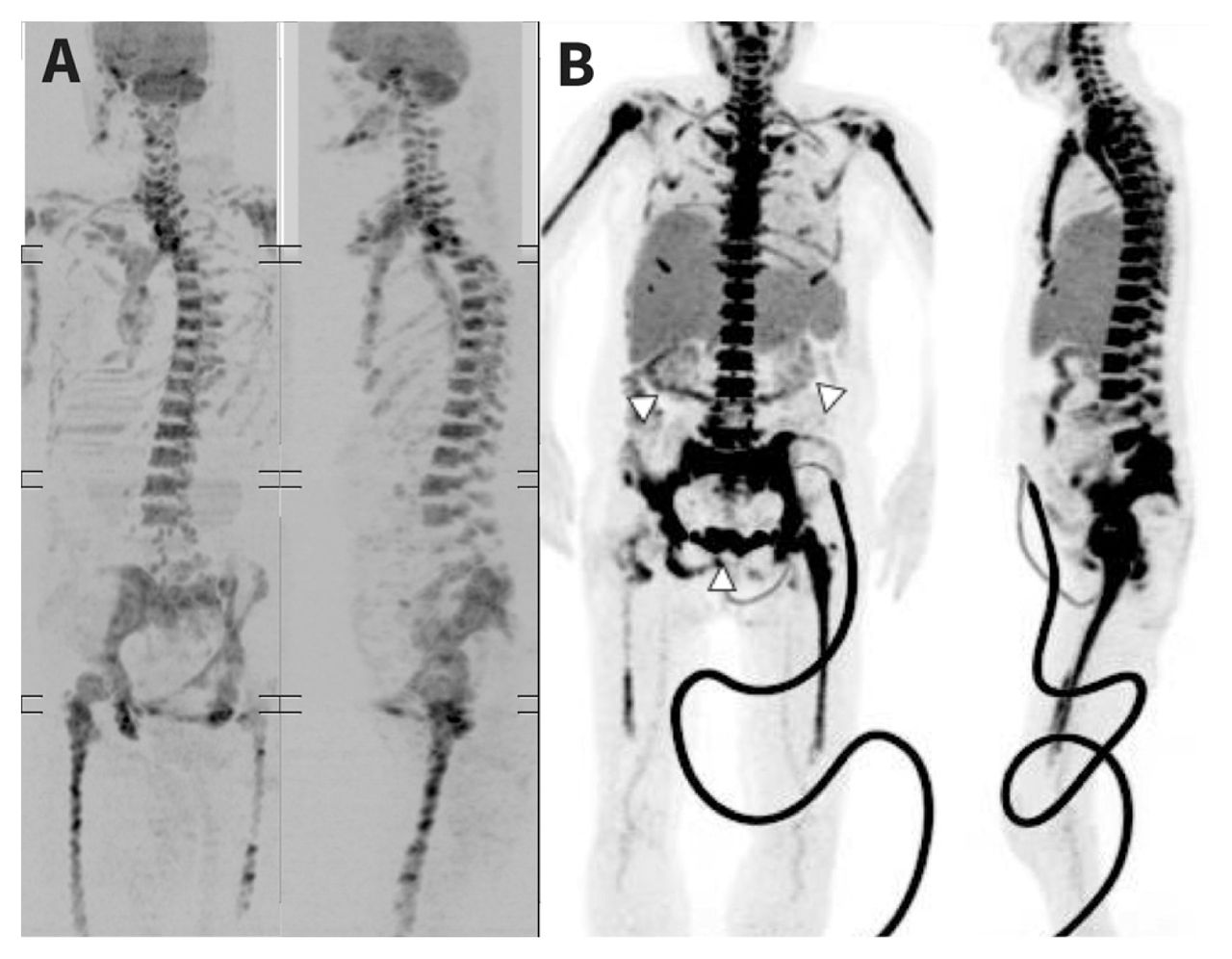
(A) Diffusion-weighted images with background body suppression (DWIBS; inverted black-and-white greyscale) in a 78-year-old woman with primary bone marrow B-cell lympoma, showing high signal intensities (in black) in the mandible, vertebral body, clavicle, sternum, humeral head, pelvis, femur, and ribs. (B) 18-Fluorodeoxyglucose positron emission tomography–computed tomography showing radionuclide uptake in the patient’s mandible, vertebral body, clavicle, sternum, humerus, part of the ribs, pelvis and femur. Hepatosplenomegaly can also be observed and decreased radionuclide uptake in the kidneys and bladder (white arrow-heads). The artificial arc on the lower body is an indwelling urinary catheter.

During the patient’s hospital stay, her serum alkaline phosphatase level (311 IU/L [reference range 106–322 IU/L]) was normal and calcium level (10.8 mg/dL [reference range 8.4–10.1 mg/dL]) was almost within the normal range. Therefore, we considered diffuse osteolytic disease unlikely and attributed the MRI findings to bone marrow disease from metastasis caused by an unknown malignant growth. We performed 18-fluorodeoxyglucose positron emission tomography–computed tomography (PET/CT) to search for the primary lesion, and the resulting images, acquired 13 days after MRI, showed diffuse uptake in the bone, similar to the DWIBS findings (Figure 1B). After a bone biopsy, we diagnosed primary bone marrow B-cell lymphoma.

The DWIBS imaging method was developed to detect tumours throughout the body using diffusion-weighted MRI.2 It has a reported detection rate equal to or better than that of bone scintigraphy for bone metastasis3 and is completed in half the time required by PET/CT and at one-sixth of the cost (in Japan). It is particularly indicated in patients who wish to avoid radiation, are breastfeeding or pregnant.

Footnotes

  • Competing interests: None declared.

  • This article has been peer reviewed.

  • The authors have obtained patient consent.

  • Funding: This study was funded by the Department of General Medicine, Osaka Medical College Hospital.

This is an Open Access article distributed in accordance with the terms of the Creative Commons Attribution (CC BY-NC-ND 4.0) licence, which permits use, distribution and reproduction in any medium, provided that the original publication is properly cited, the use is noncommercial (i.e., research or educational use), and no modifications or adaptations are made. See: https://creativecommons.org/licenses/by-nc-nd/4.0/

References

  1. ↵
    1. Osmond JD III.,
    2. Pendergrass HP,
    3. Potsaid MS
    . Accuracy of 99mTC-diphosphonate bone scans and roentgenograms in the detection of prostate, breast, and lung carcinoma metastases. Am J Roentgenol Radium Ther Nucl Med 1975;125:972–7.
    OpenUrlCrossRefPubMed
  2. ↵
    1. Takahara T,
    2. Imai Y,
    3. Yamashita T,
    4. et al
    . Diffusion weighted whole body imaging with background body signal suppression (DWIBS): technical improvement using free breathing, STIR and high-resolution 3D display. Radiat Med 2004; 22:275–82.
    OpenUrlPubMed
  3. ↵
    1. Stecco A,
    2. Lombardi M,
    3. Leva L,
    4. et al
    . Diagnostic accuracy and agreement between whole-body diffusion MRI and bone scintigraphy in detecting bone metastases. Radiol Med (Torino) 2013;118:465–75.
    OpenUrl
PreviousNext
Back to top

In this issue

Canadian Medical Association Journal: 193 (2)
CMAJ
Vol. 193, Issue 2
11 Jan 2021
  • Table of Contents
  • Index by author

Article tools

Respond to this article
Print
Download PDF
Article Alerts
To sign up for email alerts or to access your current email alerts, enter your email address below:
Email Article

Thank you for your interest in spreading the word on CMAJ.

NOTE: We only request your email address so that the person you are recommending the page to knows that you wanted them to see it, and that it is not junk mail. We do not capture any email address.

Enter multiple addresses on separate lines or separate them with commas.
“Superscan” in diffusion-weighted imaging with background body suppression magnetic resonance imaging
(Your Name) has sent you a message from CMAJ
(Your Name) thought you would like to see the CMAJ web site.
CAPTCHA
This question is for testing whether or not you are a human visitor and to prevent automated spam submissions.
Citation Tools
“Superscan” in diffusion-weighted imaging with background body suppression magnetic resonance imaging
Fumio Shimada, Miwa Misawa, Tomio Suzuki
CMAJ Jan 2021, 193 (2) E48; DOI: 10.1503/cmaj.191188

Citation Manager Formats

  • BibTeX
  • Bookends
  • EasyBib
  • EndNote (tagged)
  • EndNote 8 (xml)
  • Medlars
  • Mendeley
  • Papers
  • RefWorks Tagged
  • Ref Manager
  • RIS
  • Zotero
‍ Request Permissions
Share
“Superscan” in diffusion-weighted imaging with background body suppression magnetic resonance imaging
Fumio Shimada, Miwa Misawa, Tomio Suzuki
CMAJ Jan 2021, 193 (2) E48; DOI: 10.1503/cmaj.191188
Digg logo Reddit logo Twitter logo Facebook logo Google logo Mendeley logo
  • Tweet Widget
  • Facebook Like

Jump to section

  • Article
    • Footnotes
    • References
  • Figures & Tables
  • Responses
  • Metrics
  • PDF

Related Articles

  • Hypercaptation à l’IRM pondérée en diffusion avec suppression des signaux corporels non pertinents
  • PubMed
  • Google Scholar

Cited By...

  • No citing articles found.
  • Google Scholar

More in this TOC Section

  • Parechovirus infections in infants
  • Radiation dermatitis in a patient treated for hepatocarcinoma
  • Spontaneous bacterial peritonitis in cirrhosis
Show more Practice

Similar Articles

Collections

  • Article Types
    • Clinical Images
  • Topics
    • Cancer & oncology
    • Imaging
    • Internal medicine
    • Pain


View Latest Classified Ads

Content

  • Current issue
  • Past issues
  • Collections
  • Sections
  • Blog
  • Podcasts
  • Alerts
  • RSS
  • Early releases

Information for

  • Advertisers
  • Authors
  • Reviewers
  • CMA Members
  • CPD credits
  • Media
  • Reprint requests
  • Subscribers

About

  • General Information
  • Journal staff
  • Editorial Board
  • Advisory Panels
  • Governance Council
  • Journal Oversight
  • Careers
  • Contact
  • Copyright and Permissions
  • Accessibiity
  • CMA Civility Standards
CMAJ Group

Copyright 2023, CMA Impact Inc. or its licensors. All rights reserved. ISSN 1488-2329 (e) 0820-3946 (p)

All editorial matter in CMAJ represents the opinions of the authors and not necessarily those of the Canadian Medical Association or its subsidiaries.

To receive any of these resources in an accessible format, please contact us at CMAJ Group, 500-1410 Blair Towers Place, Ottawa ON, K1J 9B9; p: 1-888-855-2555; e: cmajgroup@cmaj.ca

Powered by HighWire