


What's your call?
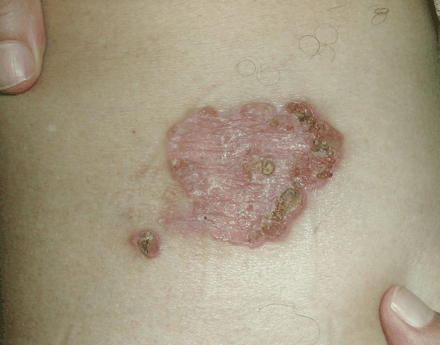
Figure. A 70-year-old otherwise healthy man presented with a 1-year history of these 2 gradually enlarging asymptomatic lesions over the right thigh.
Bowen disease is most commonly found in white patients over 60 years old. Other risk factors include chronic sun exposure, immunosuppression, arsenic exposure and cutaneous human papillomavirus (HPV) infection. HPV types 16, 18, 34 and 48 cause Bowen disease at genital sites; the role of HPV in nongenital cases of Bowen disease is less well defined. HPV types 2, 16, 34 and 35 have been rarely identified within nongenital lesions.
The lesions occur on mucous or cutaneous surfaces exposed to the sun (hands, head, neck) as a solitary, asymptomatic, sharply demarcated, scaly and erythematous plaque, measuring from a few millimetres to several centimetres in diameter (Fig. 1). The lesions may be fissured or verrucous or, rarely, pigmented. Ulceration may occur and is often a sign that invasive disease is developing. The lesion extends progressively in an annular or polycyclic pattern. The risk of progression of Bowen disease to invasive carcinoma is about 3%.1

Fig. 1: The 2 lesions (arrows) with their well-defined and erythematous areas, and central and peripheral crusts and erosions were biopsied. Histology (images are available online at www.cmaj.ca/cgi/content/full/175/7/739/DC1) demonstrated changes consistent with squamous cell carcinoma in situ, or Bowen disease, with no dermal invasion.
The differential diagnosis includes superficial basal cell carcinoma, psoriasis and eczema, which can be differentiated histopathologically.
Treatment options are broad and include surgical excision, curettage and electrodessication, cryotherapy, topical administration of 5-fluorouracil, imiquimod, photodynamic therapy and CO2 laser therapy. The choice of treatment depends on the size, location and accessibility of these therapies.
In this issue
Article tools


Jump to section
Related Articles
Cited By...
- No citing articles found.
More in this TOC Section
Similar Articles
Collections

