
Main menu

  • Home
  • Content
    • Current issue
    • Past issues
    • Early releases
    • Collections
    • Sections
    • Blog
    • Infographics & illustrations
    • Podcasts
    • COVID-19 Articles
  • Authors & Reviewers
    • Overview for authors
    • Submission guidelines
    • Submit a manuscript
    • Forms
    • Editorial process
    • Editorial policies
    • Peer review process
    • Publication fees
    • Reprint requests
    • Open access
    • Patient engagement
  • Members & Subscribers
    • Benefits for CMA Members
    • CPD Credits for Members
    • Subscribe to CMAJ Print
    • Subscription Prices
    • Obituary notices
  • Alerts
    • Email alerts
    • RSS
  • JAMC
    • À propos
    • Numéro en cours
    • Archives
    • Sections
    • Abonnement
    • Alertes
    • Trousse média 2023
  • CMAJ JOURNALS
    • CMAJ Open
    • CJS
    • JAMC
    • JPN

User menu

Search

  • Advanced search
CMAJ
  • CMAJ JOURNALS
    • CMAJ Open
    • CJS
    • JAMC
    • JPN
CMAJ

Advanced Search

  • Home
  • Content
    • Current issue
    • Past issues
    • Early releases
    • Collections
    • Sections
    • Blog
    • Infographics & illustrations
    • Podcasts
    • COVID-19 Articles
  • Authors & Reviewers
    • Overview for authors
    • Submission guidelines
    • Submit a manuscript
    • Forms
    • Editorial process
    • Editorial policies
    • Peer review process
    • Publication fees
    • Reprint requests
    • Open access
    • Patient engagement
  • Members & Subscribers
    • Benefits for CMA Members
    • CPD Credits for Members
    • Subscribe to CMAJ Print
    • Subscription Prices
    • Obituary notices
  • Alerts
    • Email alerts
    • RSS
  • JAMC
    • À propos
    • Numéro en cours
    • Archives
    • Sections
    • Abonnement
    • Alertes
    • Trousse média 2023
  • Visit CMAJ on Facebook
  • Follow CMAJ on Twitter
  • Follow CMAJ on Pinterest
  • Follow CMAJ on Youtube
  • Follow CMAJ on Instagram
Clinical Vistas

A baby with an armoured brain

Mohammed Al Wohaibi, Neville Russell and Ahmad Al Ferayan
CMAJ July 08, 2003 169 (1) 46-47;
Mohammed Al Wohaibi
  • Find this author on Google Scholar
  • Find this author on PubMed
  • Search for this author on this site
Neville Russell
  • Find this author on Google Scholar
  • Find this author on PubMed
  • Search for this author on this site
Ahmad Al Ferayan
  • Find this author on Google Scholar
  • Find this author on PubMed
  • Search for this author on this site
  • Article
  • Figures & Tables
  • Responses
  • Metrics
  • PDF
Loading

A male infant was born at term to a woman in her 20s after an uneventful pregnancy and uncomplicated vaginal delivery. He weighed 3500 g and had normal Apgar scores. A large, intact thoracolumbar myelomeningocele was present. There was no family history of neural tube defects, and no specific antenatal testing for these defects had been carried out. The myelomeningocele was associated with a complete sensorimotor paraplegia, absent anal tone, a neurogenic bladder and equinus deformities of the feet. A CT scan of the head performed within days after birth showed hydrocephalus associated with Chiari's malformation type II. At 5 days the myelomeningocele was repaired, and several days later a ventriculoperitoneal shunt was inserted.

At age 4 months the infant presented with a shunt infection, which was treated by removal of the shunt apparatus, external ventricular drainage and appropriate antibiotic therapy. At this time a CT scan showed no evidence of subdural hematomas. When the cerebrospinal fluid became sterile, a new ventriculoperitoneal shunt was inserted. At 10 months, the boy was asymptomatic with a functioning shunt. A CT scan, carried out to assess the ventricular size and the position of the shunt catheter, revealed bilateral subdural hematomas with dense calcification, particularly prominent within the inner membrane (Fig. 1). Because the boy was asymptomatic, surgery was not contemplated. He was followed regularly as an outpatient and continued to be well with normal development, but he was lost to follow-up at age 2 years.

Figure
  • Download figure
  • Open in new tab
  • Download powerpoint

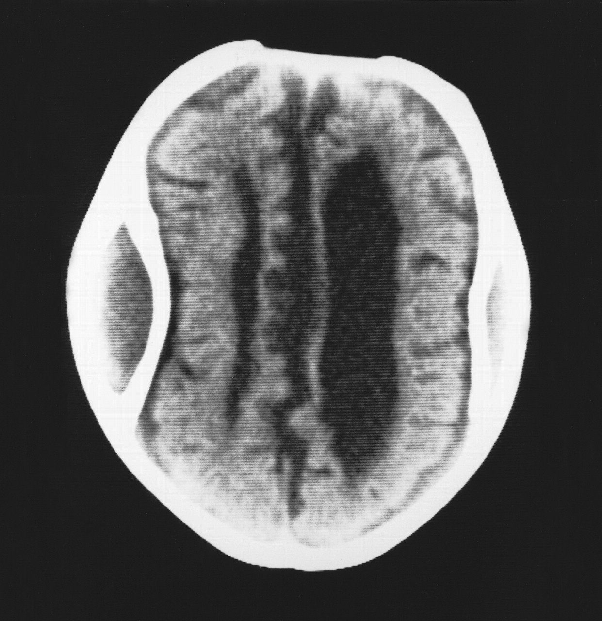
Figure 1. Photo: Image courtesy of Audio Visual Services, King Fahad National Guard Hospital

Dense calcification and even ossification of a chronic subdural hematoma is rare but has been well documented.1 When extensive, it has been likened to a shell or carapace, encasing the brain, thus the term “armoured brain.”2 The frequency with which radiologically demonstrable calcification occurs within the membranes of a chronic subdural hematoma has been reported to range from 0.3% to 2.7%.3 However, Harwood-Nash4 recorded a 10% incidence among children with chronic subdural hematomas studied by plain radiographs. Although usually seen in subdural hematomas resulting from trauma, it has also been reported in post-meningitic subdural effusions as well as subdural hematomas associated with ventricular shunting.5 The latter condition may occur because of a partial collapse of the brain when the intraventricular pressure is reduced by the shunt; this collapse results in widening of the subdural space and stretching of the bridging veins, which thereby increases the risk of hemorrhage.4 Although calcification of subdural hematomas seems more common in children, it has been reported in all age groups.1 The interval between the occurrence of the hematoma and the development of the calcification varies from 6 months to many years.1 , 5 Our patient had a normal CT scan at age 4 months; however, bilateral calcification was evident 6 months later.

The mechanism underlying the calcification remains unclear. Microscopic calcium deposits can often be found within the membranes of a chronic subdural hematoma, in its final stages of development.1 In certain cases, this mineralization may progress to more extensive calcification and even ossification.1 , 3 Etiological theories include poor circulation and delayed absorption of the hematoma fluid within the subdural space, which leads to stagnation and eventually calcification.3 In addition, an underlying metabolic abnormality such as an inherent tendency to calcification has been postulated.1 , 5 The clinical presentation varies, from patients who are asymptomatic to those with symptoms and signs of raised intracranial pressure as well as seizures and mental retardation.1 , 3 , 5

Surgical removal of the calcification is difficult.3 It should not be performed routinely; instead, it should be reserved for patients who have progressive neurological abnormalities or evidence of increased intracranial pressure.

Mohammed Al Wohaibi Neville Russell Ahmad Al Ferayan Division of Neurosurgery King Fahad National Guard Hospital Riyadh, Saudi Arabia

References

  1. 1.↵
    Afra D. Ossification of subdural hematoma: report of two cases. J Neurosurg 1961;18:393-7.
    OpenUrlPubMed
  2. 2.↵
    Ludwig B, Nix W, Lanksch W. Computed tomography of the “armored brain.” Neuroradiology 1983;25:39-43.
    OpenUrlPubMed
  3. 3.↵
    Niwa J, Nakamura T, Fujishige M, Hashi K. Removal of a large asymptomatic calcified chronic subdural hematoma. Surg Neurol 1988;30 (2):135-9.
    OpenUrlCrossRefPubMed
  4. 4.↵
    Harwood-Nash DC. Craniocerebral trauma in children. Curr Probl Radiol 1973;3:1-3.
  5. 5.↵
    Sharma RR, Mahapatra A, Pawar SJ, Sousa J, Athale SD. Symptomatic calcified subdural hematomas. Pediatr Neurosurg 1999;31:150-4.
    OpenUrlCrossRefPubMed
PreviousNext
Back to top

In this issue

Canadian Medical Association Journal: 169 (1)
CMAJ
Vol. 169, Issue 1
8 Jul 2003
  • Table of Contents
  • Index by author

Article tools

Respond to this article
Print
Download PDF
Article Alerts
To sign up for email alerts or to access your current email alerts, enter your email address below:
Email Article

Thank you for your interest in spreading the word on CMAJ.

NOTE: We only request your email address so that the person you are recommending the page to knows that you wanted them to see it, and that it is not junk mail. We do not capture any email address.

Enter multiple addresses on separate lines or separate them with commas.
A baby with an armoured brain
(Your Name) has sent you a message from CMAJ
(Your Name) thought you would like to see the CMAJ web site.
CAPTCHA
This question is for testing whether or not you are a human visitor and to prevent automated spam submissions.
Citation Tools
A baby with an armoured brain
Mohammed Al Wohaibi, Neville Russell, Ahmad Al Ferayan
CMAJ Jul 2003, 169 (1) 46-47;

Citation Manager Formats

  • BibTeX
  • Bookends
  • EasyBib
  • EndNote (tagged)
  • EndNote 8 (xml)
  • Medlars
  • Mendeley
  • Papers
  • RefWorks Tagged
  • Ref Manager
  • RIS
  • Zotero
‍ Request Permissions
Share
A baby with an armoured brain
Mohammed Al Wohaibi, Neville Russell, Ahmad Al Ferayan
CMAJ Jul 2003, 169 (1) 46-47;
Digg logo Reddit logo Twitter logo Facebook logo Google logo Mendeley logo
  • Tweet Widget
  • Facebook Like

Jump to section

  • Article
    • References
  • Figures & Tables
  • Responses
  • Metrics
  • PDF

Related Articles

  • No related articles found.
  • PubMed
  • Google Scholar

Cited By...

  • Calcified chronic subdural haematoma
  • Armoured brain
  • Armoured brain
  • Google Scholar

More in this TOC Section

  • The mystery of the broken bones
  • Elderly woman with rapidly growing, ulcerated pigmented lesion
  • Cat naps: an elderly woman with recurrent syncope
Show more Clinical Vistas

Similar Articles

Collections

  • Topics
    • Neurology


View Latest Classified Ads

Content

  • Current issue
  • Past issues
  • Collections
  • Sections
  • Blog
  • Podcasts
  • Alerts
  • RSS
  • Early releases

Information for

  • Advertisers
  • Authors
  • Reviewers
  • CMA Members
  • CPD credits
  • Media
  • Reprint requests
  • Subscribers

About

  • General Information
  • Journal staff
  • Editorial Board
  • Advisory Panels
  • Governance Council
  • Journal Oversight
  • Careers
  • Contact
  • Copyright and Permissions
  • Accessibiity
  • CMA Civility Standards
CMAJ Group

Copyright 2023, CMA Impact Inc. or its licensors. All rights reserved. ISSN 1488-2329 (e) 0820-3946 (p)

All editorial matter in CMAJ represents the opinions of the authors and not necessarily those of the Canadian Medical Association or its subsidiaries.

To receive any of these resources in an accessible format, please contact us at CMAJ Group, 500-1410 Blair Towers Place, Ottawa ON, K1J 9B9; p: 1-888-855-2555; e: cmajgroup@cmaj.ca

Powered by HighWire