
Main menu

  • Home
  • Content
    • Current issue
    • Past issues
    • Early releases
    • Collections
    • Sections
    • Blog
    • Infographics & illustrations
    • Podcasts
    • COVID-19 articles
    • Obituary notices
  • Authors & Reviewers
    • Overview for authors
    • Submission guidelines
    • Submit a manuscript
    • Forms
    • Editorial process
    • Editorial policies
    • Peer review process
    • Publication fees
    • Reprint requests
    • Open access
    • Patient engagement
  • Physicians & Subscribers
    • Benefits for Canadian physicians
    • CPD Credits for CMA Members
    • Subscribe to CMAJ Print
    • Subscription prices
    • Obituary notices
  • Alerts
    • Email alerts
    • RSS
  • JAMC
    • À propos
    • Numéro en cours
    • Archives
    • Sections
    • Abonnement
    • Alertes
    • Trousse média 2023
    • Avis de décès
  • CMAJ JOURNALS
    • CMAJ Open
    • CJS
    • JAMC
    • JPN

User menu

Search

  • Advanced search
CMAJ
  • CMAJ JOURNALS
    • CMAJ Open
    • CJS
    • JAMC
    • JPN
CMAJ

Advanced Search

  • Home
  • Content
    • Current issue
    • Past issues
    • Early releases
    • Collections
    • Sections
    • Blog
    • Infographics & illustrations
    • Podcasts
    • COVID-19 articles
    • Obituary notices
  • Authors & Reviewers
    • Overview for authors
    • Submission guidelines
    • Submit a manuscript
    • Forms
    • Editorial process
    • Editorial policies
    • Peer review process
    • Publication fees
    • Reprint requests
    • Open access
    • Patient engagement
  • Physicians & Subscribers
    • Benefits for Canadian physicians
    • CPD Credits for CMA Members
    • Subscribe to CMAJ Print
    • Subscription prices
    • Obituary notices
  • Alerts
    • Email alerts
    • RSS
  • JAMC
    • À propos
    • Numéro en cours
    • Archives
    • Sections
    • Abonnement
    • Alertes
    • Trousse média 2023
    • Avis de décès
  • Visit CMAJ on Facebook
  • Follow CMAJ on Twitter
  • Follow CMAJ on Instagram
  • Listen to CMAJ podcasts
Clinical Vistas

Elderly woman with rapidly growing, ulcerated pigmented lesion

Vincenzo de Giorgi, Daniela Massi, Serena Sestini, Barbara Alfaioli and Paolo Carli
CMAJ November 11, 2003 169 (10) 1054;
Vincenzo de Giorgi
  • Find this author on Google Scholar
  • Find this author on PubMed
  • Search for this author on this site
Daniela Massi
  • Find this author on Google Scholar
  • Find this author on PubMed
  • Search for this author on this site
Serena Sestini
  • Find this author on Google Scholar
  • Find this author on PubMed
  • Search for this author on this site
Barbara Alfaioli
  • Find this author on Google Scholar
  • Find this author on PubMed
  • Search for this author on this site
Paolo Carli
  • Find this author on Google Scholar
  • Find this author on PubMed
  • Search for this author on this site
  • Article
  • Figures & Tables
  • Responses
  • Metrics
  • PDF
Loading

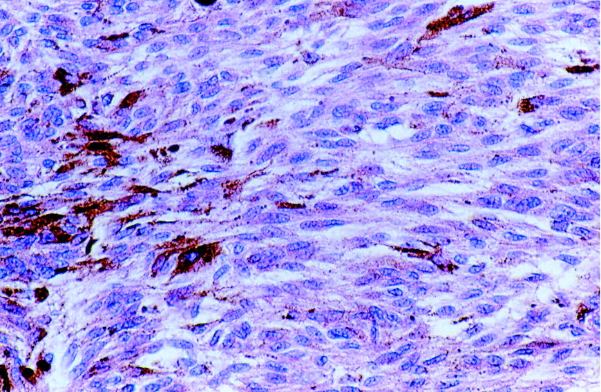
A previously well 71-year-old woman with no prior history of malignant skin cancer was referred to our dermatology clinic because of a large, pedunculated, vegetating lesion in her intergluteal region. The lesion had been present for about 30 years and had been “flat.” However, in recent years it had grown slowly and steadily and, in the few months before presentation, had rapidly increased in size. It was not painful, but there was a slight serosanguineous secretion. The lesion was dome-shaped, with a polypoid appearance, was 4 х 3.5 cm in diameter and was raised 2 cm (Fig. 1). It had a macular smooth surface and greyish-blue colour and was of a tenso-elastic consistency. There were areas of ulceration (up to 1 cm in diameter) and a hemorrhagic fundus with raised margins. The woman had no lymphadenopathy. Because of her age, the lesion's erosions and its recent evolution, a polypoid melanoma was considered, and the lesion was excised deeply with 2-cm margins. Histopathological examination showed fascicles of spindle and epithelioid cells intermingled with dendritic melanocytes and numerous melanophages consistent with a diagnosis of cellular blue nevus (Fig. 2).

Figure
  • Download figure
  • Open in new tab
  • Download powerpoint

Figure 2. Photo: Images courtesy of the Department of Dermatology, University of Florence

Figure
  • Download figure
  • Open in new tab
  • Download powerpoint

Figure 1. Photo: Images courtesy of the Department of Dermatology, University of Florence

The postoperative course was uncomplicated, and the patient was well and disease free at 1 year follow-up.

Blue nevi usually occur on the skin, although they have occasionally been observed on mucous membranes. There are essentially 2 types: common blue nevi and cellular blue nevi (CBN). The former are usually solitary (but can be multiple in number), asymptomatic, blue or black in colour, dome-shaped and less than 1 cm in diameter and, in 50% of cases, occur on or near the dorsa of the hands and feet.

CBN, the rarer variant, are usually solitary, slow growing, 1–3 cm in diameter and deep blue-black or grey in colour and have symmetrical, well-defined edges and a smooth surface. CBN are melanocytic nevi that often resemble malignant melanoma both clinically and histopathologically. They are usually situated in the superficial and middle layers of the dermis but occasionally involve the deep dermis and subcutaneous fat.1,2 Rarely are they ulcerated, but there may be areas of cysto-hemorrhagic degeneration. CBN are most often found in the sacrococcygeal and buttock regions but may also be located on the head or on the dorsa of the hands and feet. They usually are first identified in people under the age of 40, with no difference in incidence between men and women.

The differential diagnosis of CBN includes 2 clinical-histopathological forms. The first, “atypical” CBN, are not considered malignant. They have some atypical cells, often with prominent nucleoli but infrequent mitotic figures. “Malignant” CBN involve more aggressive lesions with infiltrating borders, sheet-like growth, some necrosis, frequent mitoses, nuclear pleomorphism and hyperchromasia.2,3,4

For these reasons, lesions suspected of being CBN must be surgically excised deeply into the subcutaneous tissue and with wide skin margins (at least 0.5 cm) to ensure complete removal and proper pathological distinction of simple CBN from the more aggressive subtypes and malignant melanoma.

Vincenzo de Giorgi Department of Dermatology Daniela Massi Department of Pathology and Human Oncology Serena Sestini Barbara Alfaioli Paolo Carli Department of Dermatology University of Florence Florence, Italy

References

  1. 1.↵
    Temple-Camp CR, Saxe N, King H. Benign and malignant cellular blue nevus: a clinicopathologic study of 30 cases. Am J Dermatopathol 1988;10:289-96.
    OpenUrlPubMed
  2. 2.↵
    Granter SR, McKe PH, Calonje E, Mihm MC, Busam K. Melanoma associated with blue nevus and melanoma mimicking cellular blue nevus. Am J Surg Pathol 2001;25:316-23.
    OpenUrlPubMed
  3. 3.↵
    Kang DS, Chung KY. Common blue naevus with satellite lesions: possible perivascular dissemination resulting in a clinical resemblance to malignant melanoma. Br J Dermatol 1999;141:922-5.
    OpenUrlCrossRefPubMed
  4. 4.↵
    González-Cámpora R, Galera-Davidson H, Vázquez-RamÍrez FJ, DÍaz-Cano S. Blue nevus: classical types and new related entities. A differential diagnostic review. Pathol Res Pract 1994; 190: 627-35.
    OpenUrlPubMed
PreviousNext
Back to top

In this issue

Canadian Medical Association Journal: 169 (10)
CMAJ
Vol. 169, Issue 10
11 Nov 2003
  • Table of Contents
  • Index by author

Article tools

Respond to this article
Print
Download PDF
Article Alerts
To sign up for email alerts or to access your current email alerts, enter your email address below:
Email Article

Thank you for your interest in spreading the word on CMAJ.

NOTE: We only request your email address so that the person you are recommending the page to knows that you wanted them to see it, and that it is not junk mail. We do not capture any email address.

Enter multiple addresses on separate lines or separate them with commas.
Elderly woman with rapidly growing, ulcerated pigmented lesion
(Your Name) has sent you a message from CMAJ
(Your Name) thought you would like to see the CMAJ web site.
CAPTCHA
This question is for testing whether or not you are a human visitor and to prevent automated spam submissions.
Citation Tools
Elderly woman with rapidly growing, ulcerated pigmented lesion
Vincenzo de Giorgi, Daniela Massi, Serena Sestini, Barbara Alfaioli, Paolo Carli
CMAJ Nov 2003, 169 (10) 1054;

Citation Manager Formats

  • BibTeX
  • Bookends
  • EasyBib
  • EndNote (tagged)
  • EndNote 8 (xml)
  • Medlars
  • Mendeley
  • Papers
  • RefWorks Tagged
  • Ref Manager
  • RIS
  • Zotero
‍ Request Permissions
Share
Elderly woman with rapidly growing, ulcerated pigmented lesion
Vincenzo de Giorgi, Daniela Massi, Serena Sestini, Barbara Alfaioli, Paolo Carli
CMAJ Nov 2003, 169 (10) 1054;
Digg logo Reddit logo Twitter logo Facebook logo Google logo Mendeley logo
  • Tweet Widget
  • Facebook Like

Jump to section

  • Article
    • References
  • Figures & Tables
  • Responses
  • Metrics
  • PDF

Related Articles

  • No related articles found.
  • PubMed
  • Google Scholar

Cited By...

  • No citing articles found.
  • Google Scholar

More in this TOC Section

  • The mystery of the broken bones
  • Cat naps: an elderly woman with recurrent syncope
Show more Clinical Vistas

Similar Articles

Collections

  • Topics
    • Dermatology


View Latest Classified Ads

Content

  • Current issue
  • Past issues
  • Collections
  • Sections
  • Blog
  • Podcasts
  • Alerts
  • RSS
  • Early releases

Information for

  • Advertisers
  • Authors
  • Reviewers
  • CMA Members
  • CPD credits
  • Media
  • Reprint requests
  • Subscribers

About

  • General Information
  • Journal staff
  • Editorial Board
  • Advisory Panels
  • Governance Council
  • Journal Oversight
  • Careers
  • Contact
  • Copyright and Permissions
CMAJ Group

Copyright 2023, CMA Impact Inc. or its licensors. All rights reserved. ISSN 1488-2329 (e) 0820-3946 (p)

All editorial matter in CMAJ represents the opinions of the authors and not necessarily those of the Canadian Medical Association or its subsidiaries.

To receive any of these resources in an accessible format, please contact us at CMAJ Group, 500-1410 Blair Towers Place, Ottawa ON, K1J 9B9; p: 1-888-855-2555; e: [email protected]

CMA Civility, Accessibility, Privacy


Powered by HighWire