


Article Figures & Tables
Figures
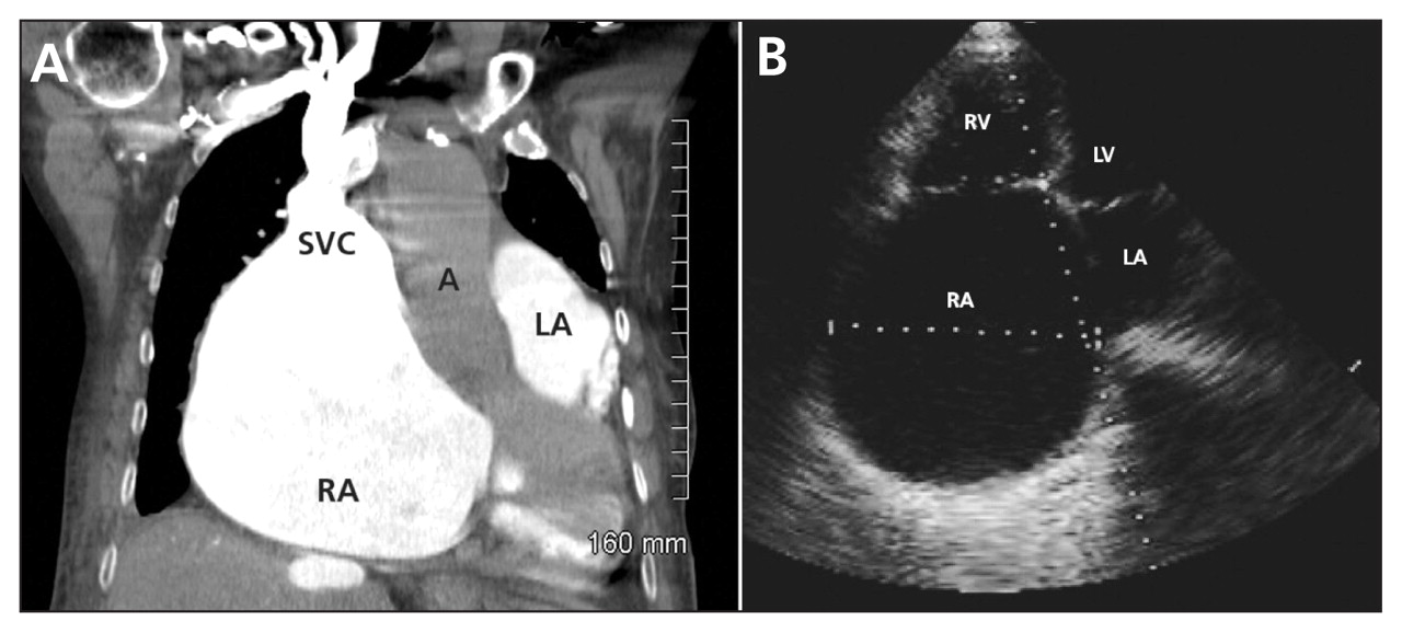
Figure 1: (A) Computed tomography scan of the chest with intravenous contrast in an 84-year-old woman showing marked dilatation of both atria and a prominent superior vena cava (SVC). (B) Echocardiogram (four-chamber apical view [enhanced for print]) showing massive enlargement of the right atrium (RA), a normal-sized left atrium (LA), which is compressed by the right atrium, and a noncoapting tricuspid valve caused by annular dilation. A = aorta, LV = left ventricle, RV = right ventricle.

In this issue

Article tools
Respond to this article


Jump to section
Related Articles
Cited By...
- No citing articles found.
More in this TOC Section
Similar Articles
Collections

