


Article Figures & Tables
Figures
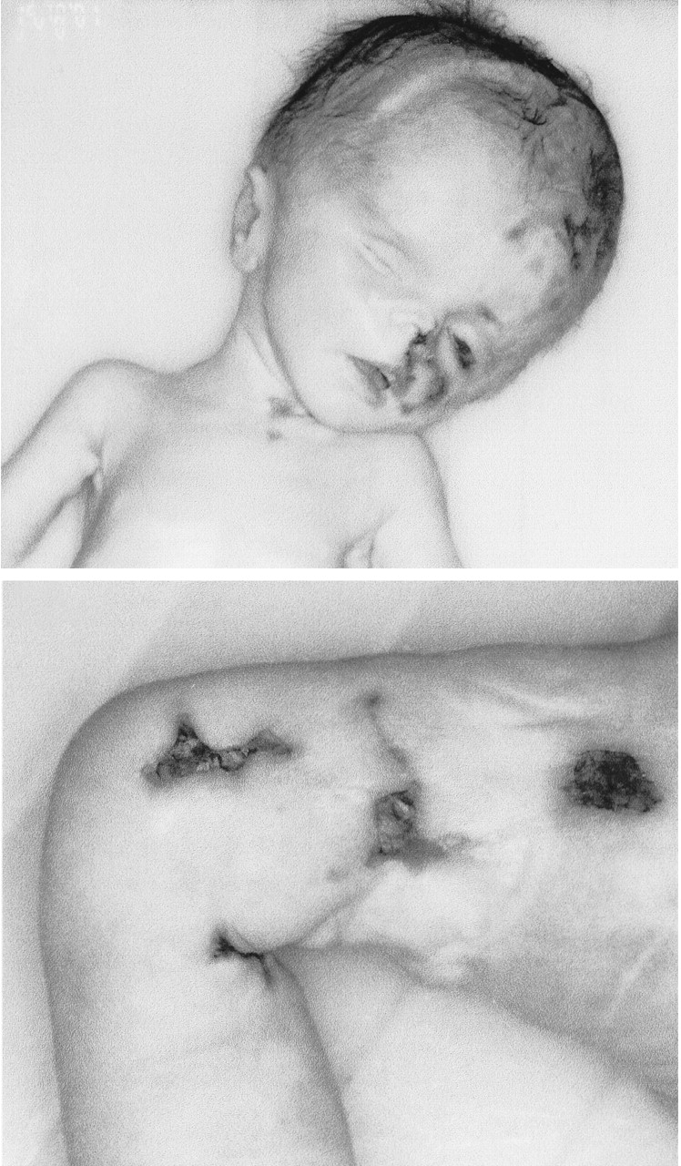
Fig. 1: Gross appearance of patient showing hydrocephalus, left-sided microphthalmia and cutaneous ulceration involving the head, neck and right lower limb.
Fig. 2: Panel A: Necrotizing encephalitis with varying degrees of gliosis and mononuclear infiltration by lymphocytes and plasma cells (hematoxylin–eosin, original magnification х10). Panel B: Focal discontinuities of the ependymal lining with subependymal infiltration by lymphocytes and gliosis (hematoxylin–eosin, original magnification х10). Panel C: Perivascular cuffing of the subependyma vessels by lymphocytes (hematoxylin–eosin, original magnification х25).

In this issue
Article tools
Respond to this article


Jump to section
Related Articles
Cited By...
More in this TOC Section
Research
Similar Articles
Collections

