
Main menu

  • Home
  • Content
    • Current issue
    • Past issues
    • Early releases
    • Collections
    • Sections
    • Blog
    • Infographics & illustrations
    • Podcasts
    • COVID-19 Articles
  • Authors & Reviewers
    • Overview for authors
    • Submission guidelines
    • Submit a manuscript
    • Forms
    • Editorial process
    • Editorial policies
    • Peer review process
    • Publication fees
    • Reprint requests
    • Open access
    • Patient engagement
  • Members & Subscribers
    • Benefits for CMA Members
    • CPD Credits for Members
    • Subscribe to CMAJ Print
    • Subscription Prices
  • Alerts
    • Email alerts
    • RSS
  • JAMC
    • À propos
    • Numéro en cours
    • Archives
    • Sections
    • Abonnement
    • Alertes
    • Trousse média 2023
  • CMAJ JOURNALS
    • CMAJ Open
    • CJS
    • JAMC
    • JPN

User menu

Search

  • Advanced search
CMAJ
  • CMAJ JOURNALS
    • CMAJ Open
    • CJS
    • JAMC
    • JPN
CMAJ

Advanced Search

  • Home
  • Content
    • Current issue
    • Past issues
    • Early releases
    • Collections
    • Sections
    • Blog
    • Infographics & illustrations
    • Podcasts
    • COVID-19 Articles
  • Authors & Reviewers
    • Overview for authors
    • Submission guidelines
    • Submit a manuscript
    • Forms
    • Editorial process
    • Editorial policies
    • Peer review process
    • Publication fees
    • Reprint requests
    • Open access
    • Patient engagement
  • Members & Subscribers
    • Benefits for CMA Members
    • CPD Credits for Members
    • Subscribe to CMAJ Print
    • Subscription Prices
  • Alerts
    • Email alerts
    • RSS
  • JAMC
    • À propos
    • Numéro en cours
    • Archives
    • Sections
    • Abonnement
    • Alertes
    • Trousse média 2023
  • Visit CMAJ on Facebook
  • Follow CMAJ on Twitter
  • Follow CMAJ on Pinterest
  • Follow CMAJ on Youtube
  • Follow CMAJ on Instagram
Practice

Rapidly enlarging scalp nodules in a 20-month-old child

Feras M. Ghazawi, Kevin Cheung and Geneviève Gavigan
CMAJ September 14, 2020 192 (37) E1066; DOI: https://doi.org/10.1503/cmaj.200314
Feras M. Ghazawi
Division of Dermatology (Ghazawi, Gavigan), University of Ottawa; Division of Plastic Surgery (Cheung), Children’s Hospital of Eastern Ontario, Ottawa, Ont.
  • Find this author on Google Scholar
  • Find this author on PubMed
  • Search for this author on this site
Kevin Cheung
Division of Dermatology (Ghazawi, Gavigan), University of Ottawa; Division of Plastic Surgery (Cheung), Children’s Hospital of Eastern Ontario, Ottawa, Ont.
  • Find this author on Google Scholar
  • Find this author on PubMed
  • Search for this author on this site
Geneviève Gavigan
Division of Dermatology (Ghazawi, Gavigan), University of Ottawa; Division of Plastic Surgery (Cheung), Children’s Hospital of Eastern Ontario, Ottawa, Ont.
  • Find this author on Google Scholar
  • Find this author on PubMed
  • Search for this author on this site
  • Article
  • Figures & Tables
  • Responses
  • Metrics
  • PDF
Loading

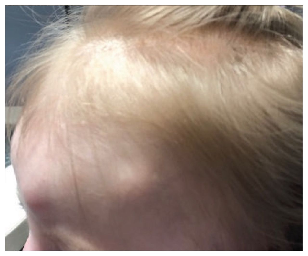
A 20-month-old girl was referred with multiple rapidly growing, painless, subcutaneous scalp nodules (Figure 1). She was otherwise well, with no history of trauma or headaches. On examination, the child appeared well. On the scalp, there were several prominent spherical, mobile and rubbery nodules with no tenderness or fluctuance. There was no overlying skin change. A history of an upper respiratory tract infection preceded the appearance of the nodules. Magnetic resonance imaging (MRI) showed 5 nonvascular and nonspecific subcutaneous lesions with no perilesional edema and no intracranial extension. Excisional biopsies of 4 lesions showed a palisaded granulomatous inflammatory process with a peripheral rim of necrobiosis, compatible with benign subcutaneous granuloma annulare, a subtype of granuloma annulare that primarily affects girls younger than 6 years.1 Granuloma annulare is usually self limited, but topical or intralesional steroids can be used for symptomatic or persistent lesions.

Figure 1:
  • Download figure
  • Open in new tab
  • Download powerpoint
Figure 1:

Scalp of a 20-month-old girl showing multiple subcutaneous nodules with no overlying skin changes.

The differential diagnosis of subcutaneous scalp nodules in babies and toddlers is broad and includes benign, infectious and malignant entities.2 Multiple rapidly growing nodules may be alarming, but benign causes include subcutaneous granuloma annulare, deep infantile hemangioma and juvenile xanthogranuloma (deep subtype). Less commonly, infections such as deep fungal and non-tuberculous mycobacterial infections may present as multiple nodules but are often associated with fever, secondary skin findings and satellite lesions. Finally, it is important to rule out diagnoses that carry high morbidity or mortality. Several malignancies may present this way, such as Langerhans cell histiocytosis, rhabdomyosarcoma, lymphoma, leukemia and metastatic neuroblastoma.

Evaluation of a baby or toddler presenting with a subcutaneous scalp nodule includes a thorough history, physical examination and investigations (e.g., imaging and histopathology) as necessary. The progression of the nodule can aid in the diagnosis; for example, whether the nodule was congenital or has grown. Constitutional symptoms may suggest malignant disease. The number, location (e.g., midline) and characteristics of the nodules on physical examination should also be taken into consideration.

Imaging studies, including ultrasonography, computed tomography and MRI, may be helpful by identifying specific features of these lesions (e.g., connecting tracts, calcification and abnormal vasculature).3 Our patient’s lesions partially resolved within a few months; no topical or interlesional steroids were used.

Footnotes

  • Competing interests: Geneviève Gavigan reports receiving honoraria from Valent, Novartis, Sanofi Genzyme, AbbVie, Lilly and L’Oréal, outside the current work. No other competing interests were declared.

  • This article has been peer reviewed.

  • The authors have obtained parental consent.

References

  1. ↵
    1. Thomas C,
    2. Morley K,
    3. Schmidt B,
    4. et al
    . Subcutaneous granuloma annulare involving the scalp. Pediatr Dermatol 2018;35:e72–3.
    OpenUrl
  2. ↵
    1. Suresh R,
    2. Sugarman J
    . Rapidly growing cutaneous nodules on the scalp. Cutis 2019;103:E16–8.
    OpenUrl
  3. ↵
    1. Rodríguez Bandera AI,
    2. Sebaratnam DF,
    3. Feito Rodríguez M,
    4. et al
    . Cutaneous ultrasound and its utility in pediatric dermatology. Part I: Lumps, bumps, and inflammatory conditions. Pediatr Dermatol 2020;37:29–39.
    OpenUrl
PreviousNext
Back to top

In this issue

Canadian Medical Association Journal: 192 (37)
CMAJ
Vol. 192, Issue 37
14 Sep 2020
  • Table of Contents
  • Index by author

Article tools

Respond to this article
Print
Download PDF
Article Alerts
To sign up for email alerts or to access your current email alerts, enter your email address below:
Email Article

Thank you for your interest in spreading the word on CMAJ.

NOTE: We only request your email address so that the person you are recommending the page to knows that you wanted them to see it, and that it is not junk mail. We do not capture any email address.

Enter multiple addresses on separate lines or separate them with commas.
Rapidly enlarging scalp nodules in a 20-month-old child
(Your Name) has sent you a message from CMAJ
(Your Name) thought you would like to see the CMAJ web site.
CAPTCHA
This question is for testing whether or not you are a human visitor and to prevent automated spam submissions.
Citation Tools
Rapidly enlarging scalp nodules in a 20-month-old child
Feras M. Ghazawi, Kevin Cheung, Geneviève Gavigan
CMAJ Sep 2020, 192 (37) E1066; DOI: 10.1503/cmaj.200314

Citation Manager Formats

  • BibTeX
  • Bookends
  • EasyBib
  • EndNote (tagged)
  • EndNote 8 (xml)
  • Medlars
  • Mendeley
  • Papers
  • RefWorks Tagged
  • Ref Manager
  • RIS
  • Zotero
‍ Request Permissions
Share
Rapidly enlarging scalp nodules in a 20-month-old child
Feras M. Ghazawi, Kevin Cheung, Geneviève Gavigan
CMAJ Sep 2020, 192 (37) E1066; DOI: 10.1503/cmaj.200314
Digg logo Reddit logo Twitter logo Facebook logo Google logo Mendeley logo
  • Tweet Widget
  • Facebook Like

Jump to section

  • Article
    • Footnotes
    • References
  • Figures & Tables
  • Responses
  • Metrics
  • PDF

Related Articles

  • No related articles found.
  • PubMed
  • Google Scholar

Cited By...

  • No citing articles found.
  • Google Scholar

More in this TOC Section

  • Parechovirus infections in infants
  • Radiation dermatitis in a patient treated for hepatocarcinoma
  • Spontaneous bacterial peritonitis in cirrhosis
Show more Practice

Similar Articles

Collections

  • Article Types
    • Clinical Images
  • Topics
    • Dermatology
    • Pediatrics


View Latest Classified Ads

Content

  • Current issue
  • Past issues
  • Collections
  • Sections
  • Blog
  • Podcasts
  • Alerts
  • RSS
  • Early releases

Information for

  • Advertisers
  • Authors
  • Reviewers
  • CMA Members
  • CPD credits
  • Media
  • Reprint requests
  • Subscribers

About

  • General Information
  • Journal staff
  • Editorial Board
  • Advisory Panels
  • Governance Council
  • Journal Oversight
  • Careers
  • Contact
  • Copyright and Permissions
  • Accessibiity
  • CMA Civility Standards
CMAJ Group

Copyright 2023, CMA Impact Inc. or its licensors. All rights reserved. ISSN 1488-2329 (e) 0820-3946 (p)

All editorial matter in CMAJ represents the opinions of the authors and not necessarily those of the Canadian Medical Association or its subsidiaries.

To receive any of these resources in an accessible format, please contact us at CMAJ Group, 500-1410 Blair Towers Place, Ottawa ON, K1J 9B9; p: 1-888-855-2555; e: cmajgroup@cmaj.ca

Powered by HighWire