


Jump to comment:
- Page navigation anchor for Authors' Response to CommentsAuthors' Response to Comments
We would like to thank Drs. Budhram, Belliveau, and Fraser for their comments. We acknowledge the errors that they identify within our article. Specifically, we agree that the patient did not have ptosis, and in fact had a lid retraction due to facial nerve palsy. Similarly, he had pontine symptoms and not lateral medullary syndrome. Finally, our paper did not clearly indicate how the patient met the dissemination in time portion of the McDonald criteria. As a result of these errors, we have made the decision to retract this article. We plan to submit a corrected version of the article shortly.
Sincerely,
Drs. Nasser, Deshauer, Caners, and ChakrabortyCompeting Interests: None declared.References
- Laila Nasser, Siobhan Deshauer, Kyla Caners, et al. A 67-year-old man with facial droop, ataxia and vertigo. CMAJ 2020;192:E626-E629.
- Page navigation anchor for Understanding application of diagnostic criteria for multiple sclerosis helps prevent misdiagnosisUnderstanding application of diagnostic criteria for multiple sclerosis helps prevent misdiagnosis
I would like to highlight several considerations regarding application of the 2017 McDonald criteria for multiple sclerosis (MS) in this case(1,2). The authors state that MS is “an atypical cause” of this patient’s lateral pontine syndrome (not lateral medullary syndrome, as they assert); if this were the case then caution should have been advised when applying the 2017 McDonald criteria, which are intended to be used primarily in patients with typical clinically isolated syndromes(2). A subacute focal brainstem presentation, however, is a typical clinically isolated syndrome(2) and use of the 2017 McDonald criteria is therefore appropriate (the patient age of onset may be atypical for MS, but the neurological presentation is not).
Regarding application of these criteria, disease dissemination both in time and space is required for the diagnosis of MS(2). In a patient such as this with one or more characteristic lesions in two or more of four areas in the CNS (i.e. periventricular, infratentorial brain and spinal cord), dissemination in space criterion has been met(2). However, the authors state this case of MS was “fulfilling McDonald criteria with the patient having 1 “attack” and objective clinical evidence of an indicative lesion on MRI in 2 areas of the central nervous system”. This explanation does not describe how the patient met criterion for dissemination in time. To meet this criterion a patient must have multiple clinical attacks, or the simultaneous pre...
Show MoreCompeting Interests: None declared.References
- 1. Laila Nasser, Siobhan Deshauer, Kyla Caners, et al. A 67-year-old man with facial droop, ataxia and vertigo. CMAJ 2020;192:E626-E629.
- 2. Alan J. Thompson, Brenda L. Banwell, Frederik Barkhof, et al. Diagnosis of multiple sclerosis: 2017 revisions of the McDonald criteria. The Lancet Neurology 2018;17(2):162-173.
- Page navigation anchor for Misinterpreted upper eyelid findingMisinterpreted upper eyelid finding
The article by Nasser and colleagues includes a common misinterpretation of a physical examination finding. This common error may lead to diagnostic confusion and also impacts triaging decisions.
The upper eyelid normally rests 1-2 mm below the superior corneoscleral limbus in most individuals (roughly at the 10 o’clock and 2 o’clock positions).
The upper eyelid position is determined by a balance between opposing forces. The retractors of the upper eyelid are the levator palpebrae superioris (innervated by the superior division of cranial nerve III) and the Muller muscle (sympathetic innervation). The protractor of the eyelids is the orbicularis oculi (innervated by cranial nerve VII).
In cranial nerve III palsy, ptosis is manifest. Ptosis also occurs with disrupted sympathetic innervation as seen in Horner syndrome.
When cranial nerve VII function is compromised, there is loss of force in the eyelid protractors and eyelid retraction may occur due to a relative unopposed force from the retractors.
With loss of force in the eyelid protractors, closure of the eye is impaired putting the individual at risk for exposure keratitis.
When misinterpreting unilateral upper eyelid retraction as upper eyelid ptosis, the implicated cranial nerve changes which impedes proper localization within the nervous system. There are of course non-neurological causes of both entities to consider as well (involutional ptosis most common; thyroid eye dise...
Show MoreCompeting Interests: None declared.References
- Laila Nasser, Siobhan Deshauer, Kyla Caners, et al. A 67-year-old man with facial droop, ataxia and vertigo. CMAJ 2020;192:E626-E629.
- Page navigation anchor for RE: Is Classification of Race Relevant in Case PresentationsRE: Is Classification of Race Relevant in Case Presentations
In the opening sentence of this fascinating paper in the Practice Section, Nasser et al inform us of this patient's "white" ethnicity. The independent contribution of race or ethnicity to the differential diagnosis of an individual presenting with a complex central nervous system syndrome is likely to be negligible. Cardiovascular, Neoplastic, metabolic, and demyelinating diseases are well described in a variety of ethnic groups. In contrast, factors such as country of origin, employment status, or educational status (that may well be be associated with ethnicity) represent variables that may be helpful in clinical problem solving.
Even if ethnicity were considered important in the formulation of a diagnosis and treatment plan, its prominent placement within the opening sentence of a patient's description gives it disproportionate emphasis. Race, especially when not self-declared by the patient, adds relatively little to the scientific impact of case presentations, while serving to support the possibility of bias in the initial assessment of a patient(1).
1. Finucane T. Virtual Mentor. 2014;16(6):423-427. doi: 10.1001/virtualmentor.2014.16.6.ecas1-1406.
Competing Interests: None declared.References
- Laila Nasser, Siobhan Deshauer, Kyla Caners, et al. A 67-year-old man with facial droop, ataxia and vertigo. CMAJ 2020;192:E626-E629.
- Page navigation anchor for Pitfalls in the recognition and localization of brainstem signsPitfalls in the recognition and localization of brainstem signs
I want to highlight a few observations about the case presented by Nasser et al(1) regarding a man with left facial droop, ataxia, and vertigo.
This patient’s findings are interesting, but I would dispute the authors’ claim that they are “pathognomonic for lateral medullary syndrome”. Perhaps the best argument for this is that this patient’s own MRI showed a lesion in the left lateral pons instead.
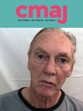
A lesion here, affecting the left vestibular nucleus, explains not only the nystagmus but also the left head tilt, which is known as an “ocular tilt reaction”. This phenomenon is not a “compensation for dysfunction of ocular alignment”, as the authors assert, but rather an intrinsically pathological response to unilateral static vestibular dysfunction.(2) In the setting of impaired graviceptive inputs from one vestibular nucleus, the brain “misperceives” gravity as pulling diagonally, and the eyes, head, and body reflexively tilt in a maladaptive effort to stay “righted”. The authors’ figure 1 actually depicts this left body tilt quite well, with the left shoulder lower than the right.(3)
I further disagree that this patient has left ptosis. The authors’ figure 3A depicts not left levator dysfunction (ptosis), but rather an overhang of redundant tissue in front of the lid (pseudoptosis). This is a common superficial mimic of ptosis called dermatochalasis, most often seen as a result of increasing age-related tissue laxity.(4) (The patient’s true left ey...
Show MoreCompeting Interests: None declared.References
- . Pitfalls in the recognition and localization of brainstem signs. 2020;:-.
- (2) Halmagyi GM, Curthoys IS, Brandt T, Dieterich M. Ocular tilt reaction: clinical sign of vestibular lesion. Acta Otolaryngol (Stockh). 1991; Suppl. 481:47-50.
- (3) Brodsky MC, Donahue SP, Vaphiades M, Brandt T. Skew deviation revisited. Surv Ophthalmol. 2006; 51: 105-128.
- (4) DeAngelis DD, Carter SR, Seiff SR. Dermatochalasis. Int Ophthalmol Clin. 2002; 42:89-101.
- Page navigation anchor for RE: A 67-year-old man with facial droop, ataxia and vertigoRE: A 67-year-old man with facial droop, ataxia and vertigo
The left head tilt may also be indicative of a right fourth nerve involvement .The fourth nerve fibers decussating as they pass through the roof of the mesencephalon. Any mention of vertical diplopia despite the nystagmus? On head tilt was a right hypertropia noted on straightening of the head or made worse on head tilt to the right side.
Competing Interests: None declared.References
- Laila Nasser, Siobhan Deshauer, Kyla Caners, et al. A 67-year-old man with facial droop, ataxia and vertigo. CMAJ 2020;192:E626-E629.
In this issue
Article tools


Jump to section
Related Articles
Cited By...
More in this TOC Section
Similar Articles

