
Main menu

  • Home
  • Content
    • Current issue
    • Past issues
    • Collections
    • Sections
    • Blog
    • Infographics & illustrations
    • Med Life with Dr. Horton
    • Podcasts
    • Videos
    • Alerts
    • RSS
    • Classified ads
  • Authors & Reviewers
    • Overview for authors
    • Submission guidelines
    • Submit a manuscript
    • Forms
    • Editorial process
    • Editorial policies
    • Peer review process
    • Publication fees
    • Reprint requests
  • CMA Members
    • Overview for members
    • Activate online account
    • Look up login
    • Earn CPD Credits
    • Members Corner
    • Print copies of CMAJ
  • Subscribers
    • General information
    • View prices
    • Activate subscription
    • Look up login
    • Manage account
    • Manage IPs
    • View Reports
  • CMAJ JOURNALS
    • CMAJ Open
    • CJS
    • JPN

User menu

  • Subscribe
  • My alerts
  • My Cart
  • Log in

Search

  • Advanced search
CMAJ
  • CMAJ JOURNALS
    • CMAJ Open
    • CJS
    • JPN
  • Subscribe
  • My alerts
  • My Cart
  • Log in
CMAJ

Advanced Search

  • Home
  • Content
    • Current issue
    • Past issues
    • Collections
    • Sections
    • Blog
    • Infographics & illustrations
    • Med Life with Dr. Horton
    • Podcasts
    • Videos
    • Alerts
    • RSS
    • Classified ads
  • Authors & Reviewers
    • Overview for authors
    • Submission guidelines
    • Submit a manuscript
    • Forms
    • Editorial process
    • Editorial policies
    • Peer review process
    • Publication fees
    • Reprint requests
  • CMA Members
    • Overview for members
    • Activate online account
    • Look up login
    • Earn CPD Credits
    • Members Corner
    • Print copies of CMAJ
  • Subscribers
    • General information
    • View prices
    • Activate subscription
    • Look up login
    • Manage account
    • Manage IPs
    • View Reports
  • Visit CMAJ on Facebook
  • Follow CMAJ on Twitter
  • Follow CMAJ on Pinterest
  • Follow CMAJ on Youtube
  • Follow CMAJ on Instagram
Practice

Japanese encephalitis

Li-Ta Keng and Lih-Yu Chang
CMAJ May 28, 2018 190 (21) E657; DOI: https://doi.org/10.1503/cmaj.171341
Li-Ta Keng
Department of Internal Medicine, National Taiwan University Hospital Hsin-Chu Branch, Hsin-Chu, Taiwan
  • Find this author on Google Scholar
  • Find this author on PubMed
  • Search for this author on this site
Lih-Yu Chang
Department of Internal Medicine, National Taiwan University Hospital Hsin-Chu Branch, Hsin-Chu, Taiwan
  • Find this author on Google Scholar
  • Find this author on PubMed
  • Search for this author on this site
  • Article
  • Figures & Tables
  • Responses
  • Metrics
  • PDF
Loading

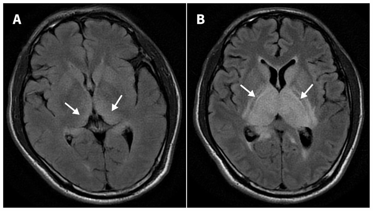
A previously healthy 52-year-old man was admitted with fever, headache and drowsiness of one day’s duration. He had neck stiffness and equivocal Kernig and Brudzinski signs. Cerebrospinal fluid showed an elevated protein level, a normal glucose concentration and lymphocytic pleocytosis. The patient’s mental status deteriorated on day 4. He became comatose and required mechanical ventilation. No intracranial hemorrhage was detected on brain computed tomography (CT). Serial brain magnetic resonance imaging (MRI) with fluid-attenuated inversion recovery sequence ( Figure 1A, on day 1, and Figure 1B, on day 5) showed progressively marked high signal intensities in the bilateral thalami (arrows), caudate nuclei and internal capsules. Diffusion-weighted MRI showed hyperintensity in the same area. There was no evidence of occlusion or high-grade stenosis of the basilar artery and main trunk of the bilateral posterior cerebral arteries. The finding was highly suggestive of Japanese encephalitis, and the diagnosis was confirmed later when the virus was detected in the cerebrospinal fluid by real-time reverse transcriptase polymerase chain reaction. Despite aggressive treatment with intravenous immunoglobulin and high-dose intravenous steroids, the patient died 30 days after admission.

Figure 1:
  • Download figure
  • Open in new tab
  • Download powerpoint
Figure 1:

(A) Brain magnetic resonance imaging (MRI) scan with fluid-attenuated inversion recovery (FLAIR) sequence on day 1 showing mildly high signal intensities in the bilateral thalami (arrows) in a 52-year-old man with Japanese encephalitis. (B) Brain MRI with FLAIR sequence on day 5 showing markedly high signal intensities in the bilateral thalami (arrows), caudate nuclei and internal capsules.

Japanese encephalitis virus, a mosquito-borne flavivirus, is the most common cause of viral encephalitis in Asia and the western Pacific rim. Although most of those infected are asymptomatic, 20%–30% of those who present with overt disease have fatal cases, and 30%–50% of survivors have neurologic, cognitive or behavioural sequelae.1 Clinical features include acute febrile illness, altered mental status, acute psychosis, seizure and acute flaccid paralysis. Thalamic abnormalities seen on imaging, such as hypodensity on CT and hyperintensity on T2-weighted MRI, have a high specificity for Japanese encephalitis in endemic areas.2 Although MRI is more sensitive than CT in detecting thalamic lesions in Japanese encephalitis,3 its diagnostic utility in the very early stage of the disease remains uncertain. Because of slowness to diagnosis, the reported mean duration between disease onset and MRI is about 10–20 days.2,3 The differential diagnosis includes metabolic diseases (e.g., hypoxic-ischemic encephalopathy, Wilson disease or Wernicke encephalopathy), vascular diseases (e.g., deep cerebral vein thrombosis or arterial occlusion), neoplasms (e.g., primary thalamic glioma) and flavivirus encephalitis other than Japanese encephalitis (e.g., West Nile fever and Murray Valley fever).4 The treatment for Japanese encephalitis remains supportive with no effective therapy.5

Footnotes

  • Competing interests: None declared.

  • This article has been peer reviewed.

  • The authors have obtained consent from the patient’s wife.

References

  1. ↵
    1. Yun SI,
    2. Lee YM
    . Japanese encephalitis: the virus and vaccines. Hum Vaccin Immunother 2014;10:263–79.
    OpenUrlCrossRefPubMed
  2. ↵
    1. Dung NM,
    2. Turtle L,
    3. Chong WK,
    4. et al
    . An evaluation of the usefulness of neuroimaging for the diagnosis of Japanese encephalitis. J Neurol 2009; 256: 2052–60.
    OpenUrlCrossRefPubMed
  3. ↵
    1. Kalita J,
    2. Misra UK
    . Comparison of CT scan and MRI findings in the diagnosis of Japanese encephalitis. J Neurol Sci 2000;174:3–8.
    OpenUrlCrossRefPubMed
  4. ↵
    1. Hegde AN,
    2. Mohan S,
    3. Lath N,
    4. et al
    . Differential diagnosis for bilateral abnormalities of the basal ganglia and thalamus. Radiographics 2011;31:5–30.
    OpenUrlCrossRefPubMed
  5. ↵
    1. Diagana M,
    2. Preux PM,
    3. Dumas M
    . Japanese encephalitis revisited. J Neurol Sci 2007;262:165–70.
    OpenUrlCrossRefPubMed
PreviousNext
Back to top

In this issue

Canadian Medical Association Journal: 190 (21)
CMAJ
Vol. 190, Issue 21
28 May 2018
  • Table of Contents
  • Index by author

Article tools

Respond to this article
Print
Download PDF
Article Alerts
To sign up for email alerts or to access your current email alerts, enter your email address below:
Email Article

Thank you for your interest in spreading the word on CMAJ.

NOTE: We only request your email address so that the person you are recommending the page to knows that you wanted them to see it, and that it is not junk mail. We do not capture any email address.

Enter multiple addresses on separate lines or separate them with commas.
Japanese encephalitis
(Your Name) has sent you a message from CMAJ
(Your Name) thought you would like to see the CMAJ web site.
Citation Tools
Japanese encephalitis
Li-Ta Keng, Lih-Yu Chang
CMAJ May 2018, 190 (21) E657; DOI: 10.1503/cmaj.171341

Citation Manager Formats

  • BibTeX
  • Bookends
  • EasyBib
  • EndNote (tagged)
  • EndNote 8 (xml)
  • Medlars
  • Mendeley
  • Papers
  • RefWorks Tagged
  • Ref Manager
  • RIS
  • Zotero
‍ Request Permissions
Share
Japanese encephalitis
Li-Ta Keng, Lih-Yu Chang
CMAJ May 2018, 190 (21) E657; DOI: 10.1503/cmaj.171341
del.icio.us logo Digg logo Reddit logo Twitter logo CiteULike logo Facebook logo Google logo Mendeley logo
  • Tweet Widget
  • Facebook Like
  • Google Plus One

Jump to section

  • Article
    • Footnotes
    • References
  • Figures & Tables
  • Responses
  • Metrics
  • PDF

Related Articles

  • No related articles found.
  • Scopus
  • PubMed
  • Google Scholar

Cited By...

  • No citing articles found.
  • Google Scholar

More in this TOC Section

  • Zollinger–Ellison syndrome
  • Benign splenic regrowth mistaken as recurrent renal cell carcinoma
  • Plastic bronchitis in a 3-year-old boy
Show more Practice

Similar Articles

Collections

  • Areas of Focus
    • Sepsis
  • Sections
    • Clinical Images
  • Topics
    • Neurology
    • Infectious diseases
    • Critical & intensive care
    • Imaging

Content

  • Current issue
  • Past issues
  • Collections
  • Sections
  • Blog
  • Podcasts
  • Videos
  • Alerts
  • RSS

Information for

  • Advertisers
  • Authors
  • CMA Members
  • Copyright and Permissions
  • Media
  • Reprint requests
  • Subscribers

About

  • General Information
  • Journal staff
  • Editorial Board
  • Governance Council
  • Journal Oversight
  • Careers
  • Contact

Copyright 2019, Joule Inc. or its licensors. All rights reserved. ISSN 1488-2329 (e) 0820-3946 (p)

All editorial matter in CMAJ represents the opinions of the authors and not necessarily those of the Canadian Medical Association or its subsidiaries.

Powered by HighWire