


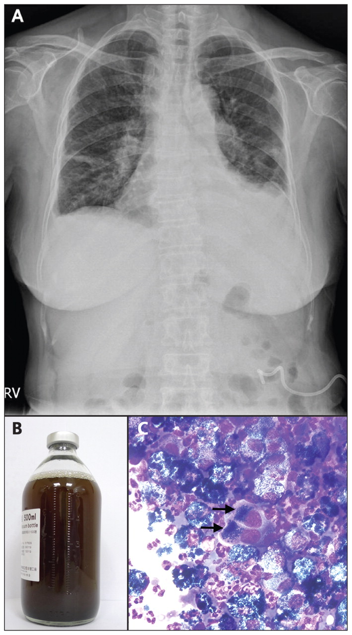
A 71-year-old woman with a history of recurrent melanoma presented with a 20-day history of left-sided chest pain. On physical examination, she was afebrile and her vital signs were normal.
Over her left lower lung field, there was dullness to percussion and she had decreased breath sounds. A chest radiograph was consistent with left pleural effusion (Figure 1A), which was confirmed by ultrasonography. Subsequent thoracentesis yielded a black fluid (Figure 1B). Cytologic examination of the fluid by means of Diff-Quick stain showed melanin pigment deposits within enlarged cells with hyperchromatic nuclei and conspicuous nucleoli (Figure 1C). The diagnosis was melanoma with left-sided malignant pleural effusion. Conservative management with the implantation of a 12-F pigtail catheter helped improve the patient’s symptoms within days and she declined further treatment of her metastatic melanoma.

Figure 1: (A) Chest radiograph of a 71-year-old woman, demonstrating a left pleural effusion. (B) Black fluid yielded from chest ultrasonography–guided thoracentesis. (C) Cytologic view of the fluid (Diff-Quick stain, original magnification ×400), showing melanin pigment deposits within enlarged cells with hyperchromatic nuclei and conspicuous nucleoli (arrows).
The appearance of pleural fluid is related to its causes. “Serous” and “blood tinged” are the most common descriptions recorded for pleural fluid at thoracentesis. 1,2 Black pleural effusions have been reported, and are usually related to infection (bacterial or fungal) or hemorrhage. 3,4 Melanoma metastasizes to the thorax in about 16% of cases, and of these, malignant pleural effusion (e.g., may be black) is observed in about 2%. 2 Metastatic melanoma has also been associated with black ascites. 5
Footnotes
-
Previously published at www.cmaj.ca
This article has been peer reviewed.
Competing interests: None declared.
In this issue
Article tools


Jump to section
Related Articles
Cited By...
- No citing articles found.
More in this TOC Section
Similar Articles

