
Main menu

  • Home
  • Content
    • Current issue
    • Past issues
    • Early releases
    • Collections
    • Sections
    • Blog
    • Infographics & illustrations
    • Podcasts
    • COVID-19 Articles
    • Obituary notices
  • Authors & Reviewers
    • Overview for authors
    • Submission guidelines
    • Submit a manuscript
    • Forms
    • Editorial process
    • Editorial policies
    • Peer review process
    • Publication fees
    • Reprint requests
    • Open access
    • Patient engagement
  • Members & Subscribers
    • Benefits for CMA Members
    • CPD Credits for Members
    • Subscribe to CMAJ Print
    • Subscription Prices
    • Obituary notices
  • Alerts
    • Email alerts
    • RSS
  • JAMC
    • À propos
    • Numéro en cours
    • Archives
    • Sections
    • Abonnement
    • Alertes
    • Trousse média 2023
    • Avis de décès
  • CMAJ JOURNALS
    • CMAJ Open
    • CJS
    • JAMC
    • JPN

User menu

Search

  • Advanced search
CMAJ
  • CMAJ JOURNALS
    • CMAJ Open
    • CJS
    • JAMC
    • JPN
CMAJ

Advanced Search

  • Home
  • Content
    • Current issue
    • Past issues
    • Early releases
    • Collections
    • Sections
    • Blog
    • Infographics & illustrations
    • Podcasts
    • COVID-19 Articles
    • Obituary notices
  • Authors & Reviewers
    • Overview for authors
    • Submission guidelines
    • Submit a manuscript
    • Forms
    • Editorial process
    • Editorial policies
    • Peer review process
    • Publication fees
    • Reprint requests
    • Open access
    • Patient engagement
  • Members & Subscribers
    • Benefits for CMA Members
    • CPD Credits for Members
    • Subscribe to CMAJ Print
    • Subscription Prices
    • Obituary notices
  • Alerts
    • Email alerts
    • RSS
  • JAMC
    • À propos
    • Numéro en cours
    • Archives
    • Sections
    • Abonnement
    • Alertes
    • Trousse média 2023
    • Avis de décès
  • Visit CMAJ on Facebook
  • Follow CMAJ on Twitter
  • Follow CMAJ on Pinterest
  • Follow CMAJ on Youtube
  • Follow CMAJ on Instagram
Practice

Office management of a palpable breast lump with aspiration

Ruth E. Heisey and David R. McCready
CMAJ April 20, 2010 182 (7) 693-696; DOI: https://doi.org/10.1503/cmaj.090416
Ruth E. Heisey
  • Find this author on Google Scholar
  • Find this author on PubMed
  • Search for this author on this site
David R. McCready
  • Find this author on Google Scholar
  • Find this author on PubMed
  • Search for this author on this site
  • Article
  • Figures & Tables
  • Related Content
  • Responses
  • Metrics
  • PDF
Loading

Case

[The following is a fictitious case based on experience with similar cases.]

Debby is a 45-year-old woman with a self-detected palpable breast lump noticed incidentally one week ago. The lump is tender and increasing in size. Her sister was diagnosed with breast cancer at age 48. Debby is terrified that this may be cancer.

About 1 in 10 women who present with a new breast lump has breast cancer. 1 The younger the age of the patient at presentation, the less likely the lump will prove to be cancer. Only 1 in 100 women with a new breast lump at age 40 or younger will have breast cancer. 2 Because 10% of malignant lesions in young women have features consistent with a fibroadenoma, 3 new palpable masses in women of any age should be thoroughly evaluated. Cysts account for about 25% 4,5 of all breast lumps and are common in premenopausal women over 35 years of age and uncommon in postmenopausal women unless they have received hormone therapy. 6 In this article, we review an approach to the initial management of palpable breast lumps and describe several techniques for breast lump aspiration in the outpatient setting.

Evidence

Most information on managing breast lumps is from cohort studies. There are several algorithms available for the diagnosis of a breast lump. 1,7–9 The 1998 consensus guideline from the Department of Surgery at the University of Toronto is particularly useful for family physicians in an office setting. 8 It was updated in 2009 by a group of experts in the management of breast lumps — including four surgical oncologists (DM), a radiologist, a pathologist, a general practitioner-oncologist (RH) and a nurse practitioner —using a review of the literature since the original algorithm was developed (Figure 1). It is applicable to all postpubescent women with a discrete palpable lump that is not suspicious for cancer.

Figure
  • Download figure
  • Open in new tab
  • Download powerpoint

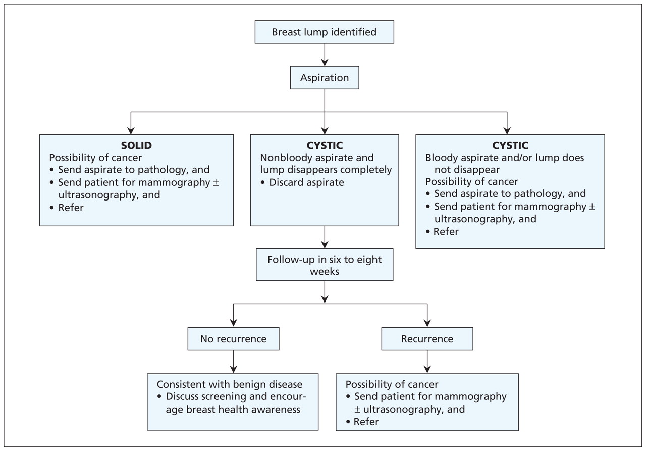
Figure 1: Consensus guideline for in-office aspiration of a discrete palpable breast lump that is not suspicious for cancer. Adapted, with permission, from Can Fam Physician 1999;45:1928. 8

Initial management

A patient presenting with a lump and features suggesting cancer (e.g., hard irregular mass fixed to the skin, palpable ipsilateral nodes or peau d’orange) should immediately be sent for mammography, ultrasonography and core biopsy, and referred to a breast surgeon.

Lumps that are not clinically suspicious for malignancy can be managed initially in the family physician’s office. The algorithm (Figure 1) advocates fine-needle aspiration of palpable lumps and assumes access to a skilled cytopathologist.

Even an experienced examiner can find it difficult to differentiate between cystic and solid lesions by palpation alone. Imaging can be useful, but the wait time may result in unnecessary anxiety for the patient. Some studies support the initial use of aspiration or ultrasound to differentiate between cystic and solid lesions. 1,9 Ultrasound is 98%–100% accurate in identifying simple cysts, but initial in-office aspiration of a cyst is therapeutic, immediately diagnostic and more cost-efficient. 1 If the physician or patient is not comfortable with breast lump aspiration, ultrasound is an alternative initial approach to determine whether the lump is cystic or solid. 1,9

Case continued

Examination showed a tender fluctuant mass measuring 2 cm in the upper outer quadrant of the right breast. Debby had no overlying skin changes or supraclavicular or axillary adenopathy.

Aspiration of a breast lump

Aspiration is a quick and straightforward initial approach for patients with a breast lump and no features suggestive of cancer. It can be done in the family physician’s office for all women with a discrete palpable mass who do not have breast implants and who are not on anticoagulants. There is no need for a local anesthetic before aspiration.

Two common approaches are described in Boxes 1 and 2. 8,10,11 Aspiration can also be practised with a model of a breast lump (Appendix 1, available at www.cmaj.ca/cgi/content/full/cmaj.090416/DC1). 12

Figure
  • Download figure
  • Open in new tab
  • Download powerpoint

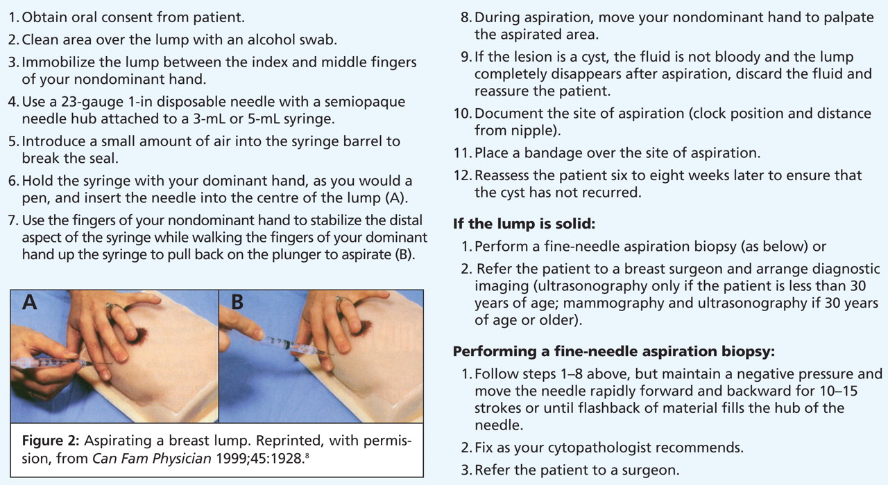
Box 1: Technique for aspirating a breast lump 8 (Figure 2)

Figure
  • Download figure
  • Open in new tab
  • Download powerpoint

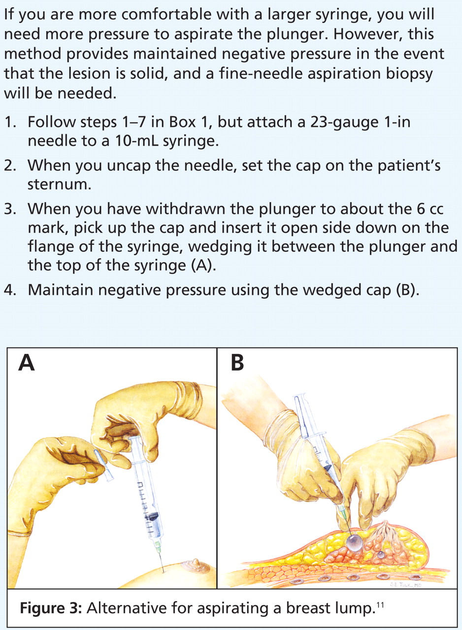
Box 2: Alternative technique (more difficult) for aspirating a breast lump 10 (Figure 3)

If the lump completely disappears with the aspiration of nonbloody fluid, the lesion is a simple cyst, and the fluid may be discarded. 13 The cyst’s location in the breast should be documented precisely (e.g., clock position and distance from the nipple).

The finding of bloody aspirate, a lump that does not completely disappear or a lump that recurs warrants referral to a surgeon. The aspirated fluid should be sent for pathologic examination. Any of these features may be associated with cancer; however, only 0.5%–1.0% of women presenting with a palpable cystic lesion will have an associated underlying neoplasm. 6,14

If the lesion is solid, a fine-needle aspiration biopsy could be performed, or the needle could be removed without further aspiration. Any specimen obtained should be fixed as per the local laboratory’s policy and sent for analysis by a cytopathologist.

Complications of aspiration include discomfort at the site of aspiration, bruising if there is inadvertent puncture of a vessel, infection, vasovagal response (which usually resolves quickly after the needle is removed) or, uncommonly, a pneumothorax. 8 If air is drawn into the syringe, immediate inspiratory and expiratory chest radiographs should be taken. Pneumothorax is unlikely if lesions close to the chest wall are moved over a rib before aspiration. 8

Aspiration does not increase the rate of false-positive results of mammography provided that the radiologist is advised about the site of aspiration. 15 If the lump proves to be cancerous, there is no evidence that a needle biopsy will cause the tumour to spread. 16 In fact, most cancers are diagnosed by a needle or core biopsy before surgery.

Further investigations

In the case of a simple cyst that resolves with aspiration, the patient should be seen six to eight weeks later to determine whether there is a recurrence of the cyst. 14 If the cyst recurs, the patient should be sent for mammography or ultrasonography, or both, and be referred to a surgeon for further assessment. If the cyst does not recur, additional tests are not necessary. This is, however, a good opportunity to ensure that screening tests are up to date.

If the lesion is solid, the patient should be sent for imaging and referred to a surgeon. If the patient is less than 30 years of age, only ultrasonography is recommended; for women 30 years of age or older, both mammography and ultrasonography should be ordered. 17 A triple assessment (examination, imaging and aspiration) to diagnose solid breast lumps ensures concordance between the clinical findings and the results of imaging and pathologic examination. 18 Although some clinicians elect to monitor palpable, presumed fibroadenomas without cytopathologic testing, this approach will miss some breast cancers in young women. In this group, most delays in the diagnosis of breast cancer result from falsely reassuring clinical findings or imaging results. 19

Case outcome

Debby’s breast lump disappeared completely after aspiration of clear fluid. She was relieved to know that the lump was only a cyst. She promised to return to the office in six weeks for a breast examination. Debby was offered mammography because she was 45 years old and had a first-degree relative with premenopausal breast cancer. However, because she had undergone mammography within the last year, there was no need for additional investigations at this time.

    Key points

  • Aspiration of a palpable breast lump allows immediate reassurance for women with breast cysts and timely investigation and referral for women with solid masses.

  • If the lump is a cyst, the aspirated fluid may be discarded provided the fluid is not bloody and the lump disappears.

  • If the lump is solid, triple assessment (clinical examination, breast imaging and fine-needle aspiration cytologic assessment) is warranted.

Footnotes

  • Previously published at www.cmaj.ca

    This article has been peer reviewed.

    Competing interests: None declared.

    Contributors: Both authors contributed to the development, writing and review of the article, and both approved the final version submitted for publication.

    Acknowledgements: The authors thank Dr. K. Bukhanov, Dr. T. Cil, Dr. W. Leong, B. Lord, Dr. M. Reedijk and Dr. B. Youngson for their assistance in updating the consensus guideline in Figure 1.

REFERENCES

  1. 1.↵
    Kerlikowske K, Smith-Bindman R, Ljung B-M, et al. Evaluation of abnormal mammography results and palpable breast abnormalities. Ann Intern Med 2003;139:274–84.
    OpenUrlCrossRefPubMed
  2. 2.↵
    Sterns EE. Age-related breast diagnosis. Can J Surg 1992;35:41–45.
    OpenUrlPubMed
  3. 3.↵
    Foxcroft LM, Evans EB, Porter AJ. The diagnosis of breast cancer in women younger than 40. Breast 2004;13:297–306.
    OpenUrlCrossRefPubMed
  4. 4.↵
    Berg WA, Campassi CI, Ioffe OB. Cystic lesions of the breast: sonographic–pathologic correlation. Radiology 2003;227:183–91.
    OpenUrlCrossRefPubMed
  5. 5.↵
    Moss HA, Britton PD, Flower CD, et al. How reliable is modern breast imaging in differentiating benign from malignant breast lesions in the symptomatic population?Clin Radiol 1999;54:676–82.
    OpenUrlCrossRefPubMed
  6. 6.↵
    Devitt JE. Breast cysts: an office-curable disorder. CMAJ 1981;124:175–6.
    OpenUrlPubMed
  7. 7.↵
    Mahoney L, Ali J, Burul C, et al. Guideline for management of breast lumps. Can J Surg 1998;41:476–7.
    OpenUrlPubMed
  8. 8.↵
    Heisey R, Mahoney L, Watson B. Management of palpable breast lumps. Consensus guideline for family physicians. Can Fam Physician 1999;45:1926–32.
    OpenUrlPubMed
  9. 9.↵
    Klein S. Evaluation of palpable breast masses. Am Fam Physician 2005;71:1731–8.
    OpenUrlPubMed
  10. 10.↵
    Henderson MA, McCready DR. A simple technique for fine needle aspiration cytology. J Am Coll Surg 1994;179:471–3.
    OpenUrlPubMed
  11. 11.↵
    Borins M. When the breasts are lumpy and painful. Patient Care Can 1996;7:63–65.
    OpenUrl
  12. 12.↵
    Delva D, Tomalty L, Payne P. Fine needle aspiration of breast lumps. Can Fam Physician 2002;48:1055–6.
    OpenUrlFREE Full Text
  13. 13.↵
    Ciatto S, Cariaggi P, Bulgaresi P. The value of routine cytologic examination of breast cyst fluids. Acta Cytol 1987;31:301–4.
    OpenUrlPubMed
  14. 14.↵
    Hamed H, Coady A, Chaudary M, et al. Follow-up of patients with breast cysts is necessary. Arch Surg 1989;124:253–5.
    OpenUrlCrossRefPubMed
  15. 15.↵
    Hindle WH, Chen EC. Accuracy of mammographic appearances after breast fine needle aspiration. Am J Obstet Gynecol 1997;176:1286–90.
    OpenUrlCrossRefPubMed
  16. 16.↵
    Peters-Engl C, Konstantiniuk P, Tausch C, et al.; Austrian Sentinel Node Biopsy Study Group. The impact of preoperative breast biopsy on the risk of sentinel lymph node metastases: analysis of 2502 cases from the Austrian Sentinel Node Biopsy Study Group. Br J Cancer 2004;91:1782–6.
    OpenUrlCrossRefPubMed
  17. 17.↵
    Parikh J. ACR appropriateness cirteria on palpable breast masses. J Am Coll Radiol 2007;4: 285–288.
    OpenUrlCrossRefPubMed
  18. 18.↵
    Kocjan G, Bourgain C, Fassina A, et al. The role of breast FNAC in diagnosis and clinical management: a survey of current practice. Cytopathology 2008;19:271–8.
    OpenUrlCrossRefPubMed
  19. 19.↵
    Brennan M, French J, Houssami N, et al. Breast cancer in young women. Aust Fam Phys 2005;34:851–5.
    OpenUrlPubMed
PreviousNext
Back to top

In this issue

Canadian Medical Association Journal: 182 (7)
CMAJ
Vol. 182, Issue 7
20 Apr 2010
  • Table of Contents
  • Index by author

Article tools

Respond to this article
Print
Download PDF
Article Alerts
To sign up for email alerts or to access your current email alerts, enter your email address below:
Email Article

Thank you for your interest in spreading the word on CMAJ.

NOTE: We only request your email address so that the person you are recommending the page to knows that you wanted them to see it, and that it is not junk mail. We do not capture any email address.

Enter multiple addresses on separate lines or separate them with commas.
Office management of a palpable breast lump with aspiration
(Your Name) has sent you a message from CMAJ
(Your Name) thought you would like to see the CMAJ web site.
CAPTCHA
This question is for testing whether or not you are a human visitor and to prevent automated spam submissions.
Citation Tools
Office management of a palpable breast lump with aspiration
Ruth E. Heisey, David R. McCready
CMAJ Apr 2010, 182 (7) 693-696; DOI: 10.1503/cmaj.090416

Citation Manager Formats

  • BibTeX
  • Bookends
  • EasyBib
  • EndNote (tagged)
  • EndNote 8 (xml)
  • Medlars
  • Mendeley
  • Papers
  • RefWorks Tagged
  • Ref Manager
  • RIS
  • Zotero
‍ Request Permissions
Share
Office management of a palpable breast lump with aspiration
Ruth E. Heisey, David R. McCready
CMAJ Apr 2010, 182 (7) 693-696; DOI: 10.1503/cmaj.090416
Digg logo Reddit logo Twitter logo Facebook logo Google logo Mendeley logo
  • Tweet Widget
  • Facebook Like

Jump to section

  • Article
    • Case
    • Evidence
    • Initial management
    • Case continued
    • Aspiration of a breast lump
    • Further investigations
    • Case outcome
    • Footnotes
    • REFERENCES
  • Figures & Tables
  • Related Content
  • Responses
  • Metrics
  • PDF

Related Articles

  • Highlights
  • PubMed
  • Google Scholar

Cited By...

  • No citing articles found.
  • Google Scholar

More in this TOC Section

  • Duodenal duplication cyst in a 61-year-old man
  • A young woman with fever and polyserositis caused by familial Mediterranean fever
  • Screening for primary aldosteronism in primary care
Show more Practice

Similar Articles

Collections

  • Topics
    • Cancer: breast


View Latest Classified Ads

Content

  • Current issue
  • Past issues
  • Collections
  • Sections
  • Blog
  • Podcasts
  • Alerts
  • RSS
  • Early releases

Information for

  • Advertisers
  • Authors
  • Reviewers
  • CMA Members
  • CPD credits
  • Media
  • Reprint requests
  • Subscribers

About

  • General Information
  • Journal staff
  • Editorial Board
  • Advisory Panels
  • Governance Council
  • Journal Oversight
  • Careers
  • Contact
  • Copyright and Permissions
  • Accessibiity
  • CMA Civility Standards
CMAJ Group

Copyright 2023, CMA Impact Inc. or its licensors. All rights reserved. ISSN 1488-2329 (e) 0820-3946 (p)

All editorial matter in CMAJ represents the opinions of the authors and not necessarily those of the Canadian Medical Association or its subsidiaries.

To receive any of these resources in an accessible format, please contact us at CMAJ Group, 500-1410 Blair Towers Place, Ottawa ON, K1J 9B9; p: 1-888-855-2555; e: cmajgroup@cmaj.ca

Powered by HighWire