


Article Figures & Tables
Figures
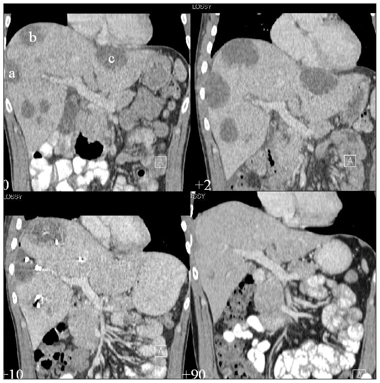
Figure 1: Coronal reconstructions of serial computed tomography scans of the abdomen in a 47-year-old man at days 0, 2, 10 and 90 show initial progression under antibiotic therapy and subsequent regression of multiple fluid collections after percutaneous drainage. Three of the abscesses are located immediately beneath the liver capsule: one laterally in the midaxillary line within segment VIII (a), another one high in the dome of the right liver, adjacent to the pleural space, again within segment VIII (b), and the third in segment II just below the pericardium (c).
Figure 2: Distribution of five drainage catheters as shown on maximum intensity projection at day 10. All catheters were removed after this CT scan because drainage had stopped.

In this issue
Article tools


Related Articles
Cited By...
- No citing articles found.
More in this TOC Section
Similar Articles

