


A 17-month-old infant with a history of prematurity presented to the pediatric dermatology clinic with multiple white nodules on the heels that were present since he was aged seven months. The patient had multiple heel sticks performed while he was in the neonatal intensive care nursery. On physical examination, only multiple white firm nodules over both heels bilaterally were observed (Figure 1).
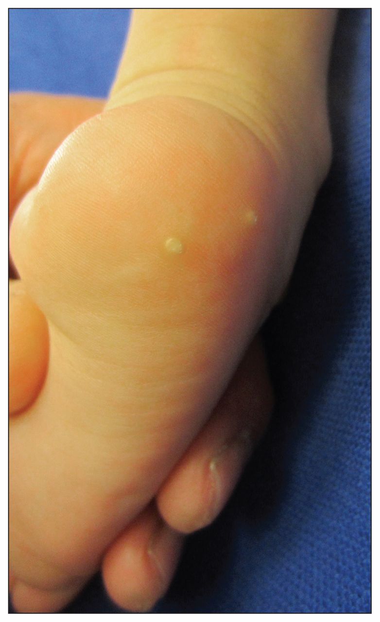
Multiple white firm nodules on the right heel of a 17-month-old infant.
Heel-stick calcinosis cutis is a form of dystrophic calcinosis cutis (calcium deposits in the skin) seen most often in infants with multiple heel sticks. Although the injury occurs in the neonatal period, the lesions do not appear until later — usually between 4 and 12 months of age.1 Lack of awareness of this condition and the prolonged delay between tissue injury and presentation may cause a delay in diagnosis.
The mechanism is thought to be secondary to the precipitation of insoluble calcium salts in the traumatized skin owing to elevated local pH from released alkaline phosphatase. An alternative possibility is that trauma to the heel may induce the formation of epidermal implantation cysts that subsequently calcify.2
The diagnosis of calcinosis cutis is usually straightforward if the condition is recognized and the clinician enquires about heel sticks in the neonatal period. Investigations are not required.
Heel-stick calcinosis cutis usully resolves spontaneously after 18–30 months.1 However, in persistent or symptomatic cases, surgical excision may be necessary and is curative.
Clinical images are chosen because they are particularly intriguing, classic or dramatic. Submissions of clear, appropriately labelled high-resolution images must be accompanied by a figure caption and the patient’s written consent for publication. A brief explanation (250 words maximum) of the educational significance of the images with minimal references is required.
Footnotes
Competing interests: None declared.
This article has been peer reviewed.
The authors have obtained patient consent.
In this issue
Article tools


Jump to section
Related Articles
Cited By...
More in this TOC Section
Similar Articles
Collections

