


Cutaneous manifestations are the most common presentation of allergic drug reactions.
Diagnosis of drug allergy is largely based on clinical history because diagnostic tests are limited.
Most patients who are labelled as having penicillin allergy can tolerate penicillins after allergy evaluation.
Cross-reactivity between cephalosporins and penicillins is rare.
Maculopapular rashes with amoxicillin are common and not an absolute contraindication for future use.
Adverse drug reactions are common both in outpatient and inpatient settings. A meta-analysis of 39 prospective studies from American hospitals that was published in 1998 reported an incidence of serious adverse drug reactions of 6.7% and fatal adverse drug reactions of 0.32%, which places these reactions around the fourth to sixth leading causes of death in the United States.3 A review published in 2005 found that adverse drug reactions affected 10%–20% of patients admitted to hospital and more than 7% of the general population.4
Health Canada has documented an increase in reported adverse drug reactions, with nearly 30 000 reports of adverse drug reactions in 2009 (up 35% from the year before).5 In addition, adverse drug reactions are more common among older patients. The Canadian Institute for Health Information (CIHI) reported that patients 65 years or older accounted for 57.6% of hospital admissions related to adverse drug reactions in Canada between 2006/07 and 2011/12, even though they accounted for only 14.2% of the Canadian population.6
Adverse drug reactions are defined by the World Health Organization (WHO) as, “all intended pharmacologic effects of a drug except therapeutic failures, intentional over-dosage, abuse of the drug, or errors in administration.”1 The WHO defines adverse drug events as “an injury resulting from medical intervention related to a drug,” which, in contrast to adverse drug reactions, also includes in its definition errors in medication use, such as overdose.2
In this review, we focus on allergic drug reactions and address key issues in diagnosis and management. The articles referenced in this review include guidelines, cohort and case–control studies, and surveys (Box 1).
Evidence used in this review
We used recent American and international practice parameters and guidelines as primary bases to inform this review, supplemented with a search for systematic reviews for supporting information. Additional clinical points or examples are based on reviews, case–control and cohort studies, and surveys. We restricted our results to articles in English. Where possible, we selected the most recent articles and the articles with the most robust level of evidence.
How are adverse drug reactions classified?
Adverse drug reactions can be classified into predictable (“type A”) and unpredictable (“type B”) reactions.7 Predictable reactions account for 80% of all adverse drug reactions; they are common, dose-dependent and caused by the pharmacologic actions of the drug.1 In contrast, unpredictable reactions are uncommon, independent of dose and unrelated to pharmacologic effects of the drug (Table 1).1 Allergic drug reactions account for about 5%–10% of adverse drug reactions overall.9 Although the term “drug allergy” has often been used exclusively for immunoglobulin E (IgE)-mediated reactions, more recently, an expert panel on drug allergy discussed whether or not the term drug allergy should also include other forms of hypersensitivity reactions that are not IgE mediated.7
What are the clinical manifestations of allergic drug reactions?
Although allergic reactions to medications can affect any organ system, cutaneous manifestations are by far the most common.8,10 A 2017 meta-analysis and systematic review of 53 studies (126 306 participants) found cutaneous manifestations to be present in 68.2% of allergic drug reactions (with anaphylactic or systemic reactions in 10.8%).11 Determining the characteristics of the cutaneous manifestation, if present, is one of the strongest diagnostic clues in a drug-induced allergic reaction.
A 2016 review on cutaneous allergic drug reactions noted that most of these eruptions are benign in nature.12 The most common cutaneous eruption is a generalized maculopapular exanthem, which accounts for up to 90% of all cutaneous eruptions caused by drugs.4,13,14 The most severe reactions are Stevens–Johnson syndrome and toxic epidermal necrolysis.8 Table 2 describes clinical features of the more common types of allergic drug reactions.
How is drug allergy diagnosed?
History
The approach to diagnosis begins with the patient’s medical history, which may identify the etiology of the reaction, identify drug allergy as a possible cause of symptoms and provide details suggesting the possible type of drug-induced allergic reaction. Table 3 provides a list of useful components of the medical history. In particular, establishing the time frame of the reaction (i.e., time of onset and its duration), the constellation of symptoms, previous exposure and underlying conditions as risk factors are essential in arriving at a diagnosis. The Naranjo Adverse Drug Reaction Probability Scale can be used, based on the patient’s history, as a validated probability scale to help determine the likelihood that the symptoms described represent an adverse drug reaction.16 This scale based on 10 questions is relatively simple to use and is frequently cited when reporting new drug allergic reactions in the literature, but it is not used commonly in clinical practice.
Laboratory tests
Laboratory investigations are supportive and not confirmatory for most allergic drug reactions. The National Institute for Health and Care Excellence recommends obtaining serum tryptase levels in the diagnosis of a potential IgE-mediated reaction, because elevated serum tryptase is relatively specific, especially if serial levels normalize, although this is based on low-quality evidence largely from observational studies.17 Serum eosinophilia supports a diagnosis of an IgE-mediated reaction, although the absence of eosinophilia does not exclude it.
Other laboratory investigations (e.g., liver enzymes, renal function, complete blood cell count) may determine involvement of internal organs, in particular with severe nonimmediate drug-induced allergic reactions. Testing for autoantibodies is useful if there is concern about vasculitis (antineutrophil cytoplasmic antibody) or drug-induced lupus (antihistone levels in systemic drug-induced lupus, and anti-Ro/SSA and anti-La/SSB for cutaneous drug-induced lupus).
Skin testing
In the diagnosis of a potential IgE-mediated reaction, validated skin testing reagents exist only for penicillin and not for any of the other low-molecular-weight drugs.1,8 Several international guidelines, including the American Academy of Allergy, Asthma and Immunology guideline, recommend skin testing (a combination of skin prick testing and then intradermal testing) with the penicillin reagents because of its high negative predictive value.8,18 This is followed by an allergist-administered oral challenge — usually of amoxicillin or penicillin in children — to prove tolerance in patients who have negative skin testing.19
The risk of or reacquiring a penicillin allergy is low after negative penicillin testing. Patients have been reported to tolerate both oral doses of penicillin1,8 and, according to a recent retrospective review, repeated intravenous penicillin without immediate hypersensitivity reactions.20
In vitro serum-specific IgE assays are available for some common antibiotics; however, their sensitivity and specificity are not well described or validated,8 although studies have found a higher specificity (90% or more) than sensitivity (29% to 68%).1,8,21 Some guidelines do recommend the inclusion of serum-specific IgE testing if skin testing is negative despite a convincing reaction history.18
The basophil activation test, which looks at in vitro basophilic stimulation with an allergen and subsequent CD63 or CD203c expression, is being reported increasingly because this test shows promise in the diagnosis of IgE-mediated drug allergy, but it is not available routinely at this time.22

Maculopapular exanthema in drug reaction with eosinophilia and systemic symptoms.

Contact dermatitis on the right shoulder.
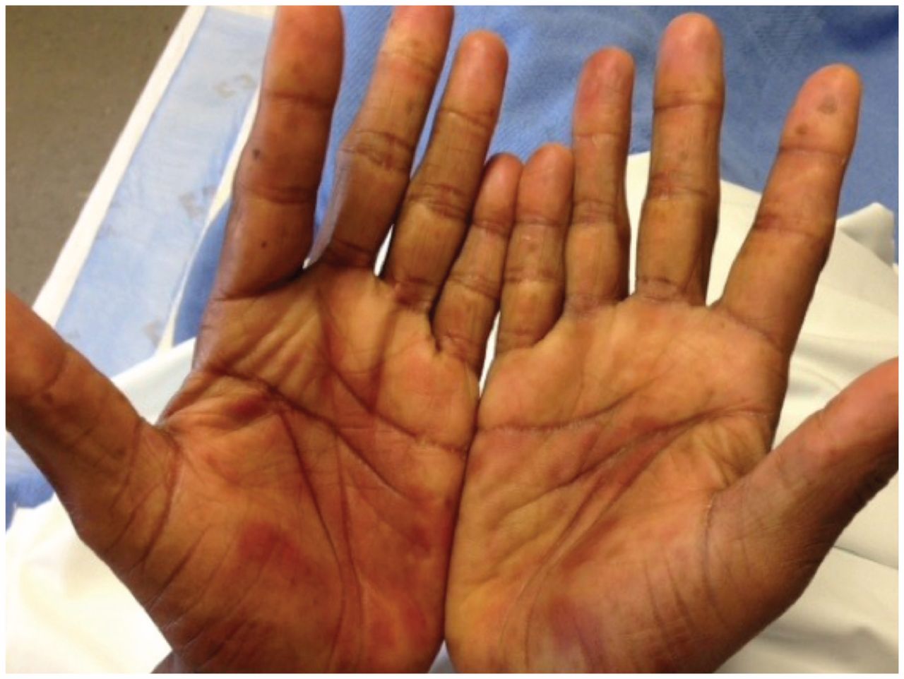
Fixed drug eruption on the palms.
For some nonimmediate reactions, in particular contact dermatitis, fixed drug eruption and maculopapular exanthem, skin patch testing (i.e., placing the allergen on the back at a nonirritating concentration under an aluminum disk) has been reported.1,23 In North America, it is not used commonly in the more severe nonimmediate drug reactions, such as Stevens–Johnson syndrome, toxic epidermal necrolysis or drug reaction with eosinophilia and systemic symptoms.8 However, in Europe, both patch testing and delayed intradermal testing (i.e., the result is read days later) are used for nonimmediate reactions, including severe reactions.24,25 A recent retrospective review of patch testing that included 260 patients who received treatment in a European dermatology clinic found that patch testing was safe and specific, even for severe nonimmediate reactions.26
Drug challenge
In most cases of drug allergy, validated skin or laboratory tests are not available. For patients in whom the likelihood of drug allergy is deemed low (e.g., remote reaction, benign rash), a drug challenge can be performed by an allergist. If the history is not indicative of an allergy (e.g., headache), a full dose can be administered to confirm tolerance. In most circumstances, a graded challenge is performed, with the assistance of an allergist, that often involves the administration of two graded sub-therapeutic doses of the medication to the patient, with monitoring for an allergic reaction. A US guideline noted that drug challenges are contraindicated if the history is consistent with a severe drug reaction, such as Stevens–Johnson syndrome, toxic epidermal necrolysis or drug reaction with eosinophilia and systemic symptoms.1
In contrast, induction of drug tolerance (e.g., drug desensitization) involves providing increasing incremental doses of the medication to the patient over a period of hours to days, using different procedures based on the hypothesized mechanism of the reaction. Induction of drug tolerance does modify the immune response to the medication temporarily while the patient remains on the medication. It effectively modifies the immune response to the medication, even though there is an underlying allergy. This procedure can be used for both IgE-and non-IgE–mediated drug allergic reactions to a variety of drugs, including antibiotics, chemotherapeutics and biologic agents (e.g., penicillin, acetylsalicylic acid [ASA] and allopurinol), but it is not used for patients with a history of a severe drug reaction, such as Stevens–Johnson syndrome, toxic epidermal necrolysis or drug reaction with eosinophilia and systemic symptoms.1
Which common drugs are associated with allergic reactions?
β-Lactam antibiotics
About 10% of the population in predominantly developed countries is thought to be penicillin allergic,1,27 but 90% or more are able to tolerate penicillin after allergy evaluation.1,28 In 2016, a systematic review and meta-analysis of 14 studies reported a low prevalence of IgE-mediated drug allergy to β-lactam antibiotics of 2.84% (95% confidence interval [CI] 1.77%–3.91%), with a higher prevalence among adults (7.78%, 95% CI 6.53%–9.04%) than among children (1.98%, 95% CI 1.35%–2.60%).29 In a 2016 Canadian retrospective chart review involving 306 patients in primary care, β-lactam allergy was ruled out in 96.1% (95% CI 93.2%–97.5%) of patients after β-lactam allergy evaluation.30
Some studies have suggested that the rate of confirmed penicillin allergy is decreasing.31,32 Recent initiatives have suggested the “de-labelling” of patients erroneously diagnosed with penicillin allergy,33 including one from Choosing Wisely Canada,34 and have noted that erroneous labelling is associated with broad-spectrum antibiotic use,1,19 increased antibiotic resistance35 and unnecessary health care costs.19,36
Penicillin
The most common drug allergic reaction to penicillin is a cutaneous reaction — either macular, morbilliform or urticarial.1 Penicillin undergoes spontaneous conversion to reactive intermediates under physiologic conditions. Most degrade to the penicilloyl moiety (major determinant) and the remainder degrade into several other moieties (minor determinants).8 Skin testing with penicillin reagents has a high negative predictive value in the diagnosis of IgE-mediated penicillin allergy, with an oral challenge by an allergist as a confirmatory step if negative.
Recent studies have shown that as many as 98% of patients with a history of penicillin allergy are found to have negative penicillin skin tests and will tolerate penicillins.36 Re-evaluation is suggested even in those with confirmed (based on skin testing or oral challenge) penicillin allergy. An evaluation conducted in a pediatric emergency department that involved 100 children with a history of penicillin allergy found that 100% (95% CI 96.4%–100%) of these children with low-risk symptoms had negative results for allergy testing (skin testing and drug provocation test).37 Evaluation is especially useful if the reaction occurred more than 5 to 10 years ago, because there is a high rate of resolution for penicillin allergy.38–40 For example, a retrospective study involving 740 patients with a history of β-lactam allergy found that 93% of these patients had a positive result for skin testing if the reaction was in the past year; this decreased to 22% of patients with a positive test result if they were evaluated 10 or more years after their clinical reaction.38
Amoxicillin and cephalosporins
Amoxicillin and ampicillin are associated with a delayed (type 4) maculopapular rash in 5%–10% of patients, and in 100% of patients with co-existing Epstein–Barr virus.8 These amoxicillin reactions are not life-threatening and not an absolute contraindication to future amoxicillin or ampicillin use.
Although cephalosporins can also cause acute allergic reactions, overall the reaction rate is about 10-fold lower than for penicillin.1 Cross reactivity between cephalosporins and penicillin is thought to be very low.41 The Canadian Pediatric Society’s guideline on otitis media notes that children with a history of reactivity to penicillin or amoxicillin can safely be prescribed second-or third-generation cephalosporins as long as the previous reaction was not life-threatening.42 A 2016 review also reported that avoidance of cephalosporins in patients with amoxicillin or penicillin allergy could result in substantial morbidity, and concluded that there was “ample evidence to allow the safe use of cephalosporins in patients with isolated confirmed penicillin or amoxicillin allergy.”43
Amoxicillin and cephalosporins contain “R” side chains in addition to the β-lactam ring, which may be allergenic. Sensitization to the β-lactam portion of penicillin would result in sensitization to all β-lactam antibiotics; in contrast, sensitization to the R side chain would lead to tolerance of most β-lactam antibiotics (except those with a common side chain). For example, amoxicillin shares an identical R side chain with cefprozil; ampicillin with cefaclor and cephalexin; and ceftriaxone with cefotaxime. For cephalosporins, if an acute allergic reaction does occur, it is often directed at the R-group side chain instead of the common β-lactam ring.8
Although skin testing has not been validated for β-lactams other than penicillin, it can still have some utility if there is a history consistent with an IgE-mediated reaction — patients with negative results for penicillin skin tests can safely receive β-lactam antibiotics, and patients with confirmed penicillin allergy usually tolerate carbapenems and aztreonam.44 Skin testing reagents have been developed for amoxicillin and cephalosporins; however, their negative predictive value has not been validated.1 If patients with a history of a reaction to amoxicillin have negative results for skin testing to the penicillin reagents, an oral challenge to amoxicillin is often considered by an allergist to rule out definitively IgE-mediated allergy. A recent Canadian cohort study also suggested that a graded oral challenge alone in an allergy clinic may be an effective diagnostic test for amoxicillin allergy in children finding that among 818 children with suspected amoxicillin allergy, a graded oral challenge was both safe and accurate. Almost all children (94.1%) tolerated the oral challenge, and the reactions when present were mild.45
The approach to administration of cephalosporins or penicillin in the context of an allergic reaction is outlined in Table 4, as suggested by the Joint Task Force on Practice Parameters.1
Administration of penicillin or cephalosporins in the context of a previous reaction1
Nonsteroidal anti-inflammatory drugs
Nonsteroidal anti-inflammatory drugs (NSAIDs) are used commonly in North America and can cause different reactions that are either allergic in nature or, more commonly, nonimmune (and related to cyclooxygenase-1 [COX-1] inhibition) (Table 5). A retrospective review involving all adult patients in an American health care system who were prescribed NSAIDs over an eight-year period reported that 17% of those patients had an adverse drug reaction, of which 18.3% were allergic.46 The common types of NSAID-induced reactions are NSAID-exacerbated respiratory disease, single–NSAID-induced anaphylaxis or urticaria/angioedema (which could be NSAID-exacerbated, NSAID-induced or single–NSAID-induced) (Table 4).47,48 Delayed reactions, such as Stevens–Johnson syndrome, delayed maculopapular rash or fixed drug eruptions, are also possible with NSAIDs.
Allergic reactions induced by nonsteroidal anti-inflammatory drugs47
Nonsteroidal anti-inflammatory drug–exacerbated respiratory disease presents with upper and lower respiratory symptoms within three hours after NSAID ingestion, mostly in adult patients with a history of underlying asthma and rhinosinusitis.47 It is related to COX-1 inhibition and is diagnosed with an oral provocation test. Treatment is avoidance of COX-1 inhibitors (COX-2 inhibitors are usually safe); if asthma or rhinosinusitis is refractory to medical and surgical therapy, ASA induction of tolerance followed by ASA therapy can be considered as well.1
Patients who present with cutaneous symptoms after NSAID exposure may have one of three conditions: NSAID-exacerbated cutaneous disease, NSAID-induced urticaria/angioedema or single-NSAID–induced urticaria/angioedema or anaphalaxis.47 All of these conditions present with angioedema/urticaria; however, the time frame differs slightly: NSAID-exacerbated cutaneous disease and NSAID-induced urticaria/angioedema can present up to several hours after NSAID ingestion (although presentation is often immediate), and single-NSAID–induced urticaria/angioedema or anaphalaxis presentation is uniformly immediate. In addition, NSAID-exacerbated cutaneous disease presents in patients with a history of chronic urticaria, and the pathophysiology differs between these conditions (Table 4). Distinguishing between these conditions by drug provocation testing (to both the implicated NSAID and a chemically unrelated NSAID) is beneficial as a means of differentiating the conditions and predicting the extent of necessary NSAID avoidance according to a 2013 review.47 For NSAID-exacerbated cutaneous disease and NSAID-induced urticaria/angioedema, all COX-1 inhibitors should be avoided (COX-2 inhibitors are usually safe). For single-NSAID–induced urticaria/angioedema or anaphalaxis, only the implicated NSAID and chemically related NSAIDs must be avoided.1,47 For all of these conditions, COX-2 inhibitors are largely well tolerated.1,47
Conclusion
Although adverse drug reactions are common, allergic reactions are uncommon. Cutaneous manifestations are the most common clinical manifestation of an allergic drug reaction. Diagnosis largely relies on medical history, because there are few standardized tests in the diagnosis of drug allergy, with the exception of skin testing for penicillin. However, evaluation of patients labelled as allergic remains an important public health goal because mislabelling can have health consequences, such as increased morbidity and public health costs.
Footnotes
Competing interests: Elissa Abrams has received an unrestricted educational grant from Novartis, outside the submitted work. No other competing interests were declared.
This article has been peer reviewed.
References
In this issue
Article tools


Jump to section
Related Articles
Cited By...
- No citing articles found.
More in this TOC Section
Similar Articles

