
Main menu

  • Home
  • COVID-19
    • Articles & podcasts
    • Blog posts
    • Collection
    • News
  • Content
    • Current issue
    • Past issues
    • Early releases
    • Collections
    • Sections
    • Blog
    • Infographics & illustrations
    • Podcasts
    • Classified ads
  • Authors
    • Overview for authors
    • Submission guidelines
    • Submit a manuscript
    • Forms
    • Editorial process
    • Editorial policies
    • Peer review process
    • Publication fees
    • Reprint requests
  • CMA Members
    • Overview for members
    • Earn CPD Credits
    • Print copies of CMAJ
    • Career Ad Discount
  • Subscribers
    • General information
    • View prices
  • Alerts
    • Email alerts
    • RSS
  • JAMC
    • À propos
    • Numéro en cours
    • Archives
    • Sections
    • Abonnement
    • Alertes
  • CMAJ JOURNALS
    • CMAJ Open
    • CJS
    • JAMC
    • JPN

User menu

Search

  • Advanced search
CMAJ
  • CMAJ JOURNALS
    • CMAJ Open
    • CJS
    • JAMC
    • JPN
CMAJ

Advanced Search

  • Home
  • COVID-19
    • Articles & podcasts
    • Blog posts
    • Collection
    • News
  • Content
    • Current issue
    • Past issues
    • Early releases
    • Collections
    • Sections
    • Blog
    • Infographics & illustrations
    • Podcasts
    • Classified ads
  • Authors
    • Overview for authors
    • Submission guidelines
    • Submit a manuscript
    • Forms
    • Editorial process
    • Editorial policies
    • Peer review process
    • Publication fees
    • Reprint requests
  • CMA Members
    • Overview for members
    • Earn CPD Credits
    • Print copies of CMAJ
    • Career Ad Discount
  • Subscribers
    • General information
    • View prices
  • Alerts
    • Email alerts
    • RSS
  • JAMC
    • À propos
    • Numéro en cours
    • Archives
    • Sections
    • Abonnement
    • Alertes
  • Visit CMAJ on Facebook
  • Follow CMAJ on Twitter
  • Follow CMAJ on Pinterest
  • Follow CMAJ on Youtube
  • Follow CMAJ on Instagram
Practice

Hereditary hemorrhagic telangiectasia: fatigue and dyspnea

Farzan Irani and Rahil Kasmani
CMAJ April 14, 2009 180 (8) 839; DOI: https://doi.org/10.1503/cmaj.081212
Farzan Irani
  • Find this author on Google Scholar
  • Find this author on PubMed
  • Search for this author on this site
Rahil Kasmani
  • Find this author on Google Scholar
  • Find this author on PubMed
  • Search for this author on this site

A 49-year-old man presented to the emergency department with a 2-week history of progressive fatigue and dyspnea, as well as recurrent epistaxis. Laboratory investigations revealed severe hypochromic microcytic anemia, with a hemoglobin level of 48 (normal 120–140) g/L. He reported that his mother and brothers also had recurrent nosebleeds.

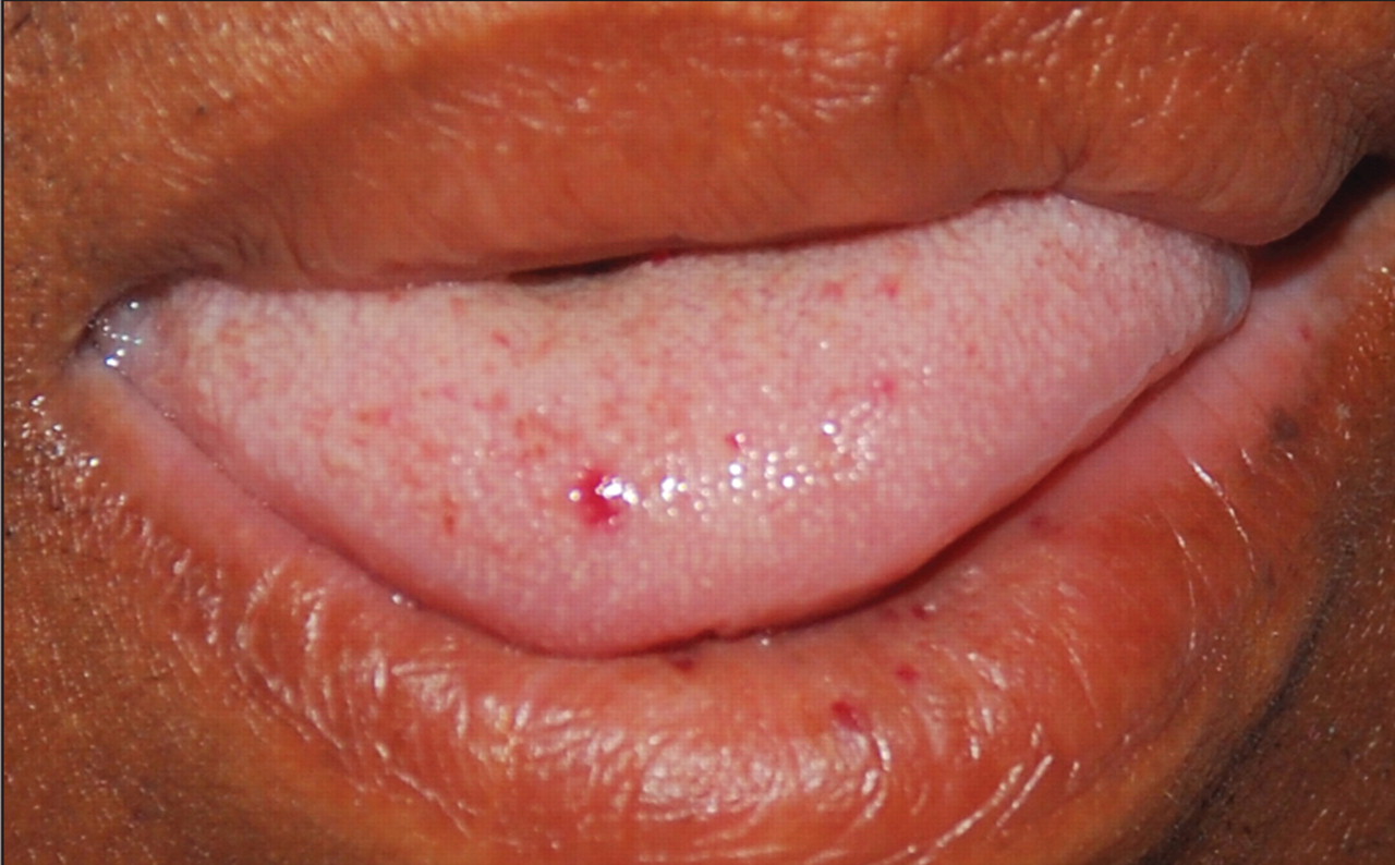
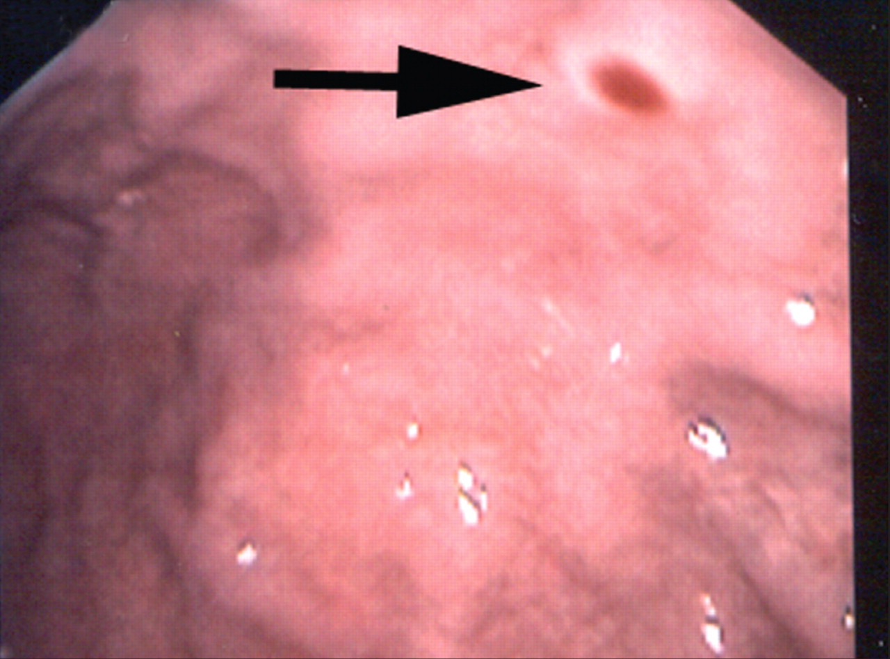
We observed multiple telangiectasias on the patient’s lower lip (Figure 1), tongue and palate. Nasal endoscopy showed active bilateral bleeding from the septum with crusting and erosions. No septal telangiectasias were found. Upper gastrointestinal endoscopy showed small nonbleeding antral and duodenal arteriovenous malformations (Figure 2). The results of a colonoscopic examination were normal.

Figure1
  • Download figure
  • Open in new tab
  • Download powerpoint

Figure 1: The patient had multiple telangiectasias on his tongue and lower lip.

Figure2
  • Download figure
  • Open in new tab
  • Download powerpoint

Figure 2: A gastric arteriovenous malformation surrounded by an anemic halo (arrow).

A computed tomography (CT) scan of his chest showed multiple arteriovenous malformations, with the largest in the anterior left lower lobe. A CT scan of the patient’s abdomen showed a large irregular arteriovenous malformation in the posterior segment of the right lobe of his liver (Figure 3), and a few other small malformations.

Figure3
  • Download figure
  • Open in new tab
  • Download powerpoint

Figure 3:(A) A computed tomography scan of the patient’s abdomen (coronal view) showing a large hepatic arteriovenous malformation (arrow). (B) A 3-dimensional reconstructed image of the patient’s liver showing a large arteriovenous malformation (arrow).

Hereditary hemorrhagic telangiectasia was diagnosed based on the patient’s recurrent epistaxis, multiple mucocutaneous telangiectasias, visceral arteriovenous malformations and family history.1 Financial constraints prevented genetic testing. Because our patient did not report headaches or other neurological symptoms, we did not perform cerebral imaging.

Neither repeat endoscopy nor nasal surgery was performed, and his nosebleed was controlled with anterior nasal packing only. A blood transfusion resulted in improvement of his symptoms. The patient’s care was continued at an outpatient hematology clinic.

See primer by Grand’Maison, page 833, case by Manawadu and colleagues, page 836, and clincial image by Nanda and Bhatt, page 838

Footnotes

  • Acknowledgement: We thank Ms. Joyce Moses BSRT for her invaluable assistance in the 3-dimensional image reconstructions.

REFERENCE

  1. 1.
    Shovlin CL, Guttmacher AE, Buscarini E, et al. Diagnostic criteria for hereditary hemorrhagic telangiectasia (Rendu–Osler–Weber syndrome). Am J Med Genet 2000;91:66–7.

Content

  • Current issue
  • Past issues
  • Collections
  • Sections
  • Blog
  • Podcasts
  • Alerts
  • RSS
  • Early releases

Information for

  • Advertisers
  • Authors
  • Reviewers
  • CMA Members
  • Media
  • Reprint requests
  • Subscribers

About

  • General Information
  • Journal staff
  • Editorial Board
  • Governance Council
  • Journal Oversight
  • Careers
  • Contact
  • Copyright and Permissions

Copyright 2021, Joule Inc. or its licensors. All rights reserved. ISSN 1488-2329 (e) 0820-3946 (p)

All editorial matter in CMAJ represents the opinions of the authors and not necessarily those of the Canadian Medical Association or its subsidiaries.

Powered by HighWire