


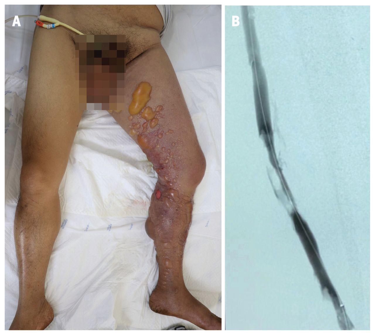
A 57-year-old man presented to the emergency department with a 2-day history of progressive severe pain and swelling from his left ankle extending to the scrotum. He was hypertensive, tachypneic and tachycardic. He had been taking nifedipine and metoprolol for hypertension. On examination, he had a blistering eruption (Figure 1A) and a weak left dorsalis pedis pulse; he was unable to tolerate sensory and motor examinations. He had elevated serum D-dimer of 6.85 (normal 0.00–0.55) mg/L fibrinogen-equivalent units and creatinine kinase of 2975 (normal 25–200) U/L, with leukocytosis of 17.42 (normal 4.00–10.00) × 109/L.

(A) Phlegmasia cerulea dolens over the medial and posterior aspects of the left leg of a 57-year-old man, which extended to his medial thigh. The leg showed prominent swelling and many well-defined blisters of variable sizes over a diffuse cyanotic background, including a mix of tense and flaccid blisters with erosions. The right leg is normal in comparison. (B) A venogram showed multiple intraluminal contrast filling defects, confirming the diagnosis of deep vein thrombosis.
Our differential diagnoses included deep venous thrombosis (DVT) and cellulitis, with possible compartment syndrome. Duplex ultrasonography showed a massive iliofemoral DVT, which was confirmed with a venogram (Figure 1B). We diagnosed phlegmasia cerulea dolens and immediately treated the patient with endovascular urokinase and mechanical thrombus removal. We discussed iIliac venous stenting, which improves vessel patency and lowers compression to prevent future episodes, but the patient declined owing to cost. Looking for a cause of his DVT, we ordered chest computed tomography, which showed a left lower lobe mass. Biopsy showed squamous cell lung cancer. His skin lesions and leg edema resolved after 3-month treatment with rivaroxaban, anlotinib and radiotherapy for his lung cancer.
Phlegmasia cerulea dolens is an uncommon condition whereby a massive DVT causes venous drainage obstruction, most often among patients aged 40–50 years.1 Risk factors include malignant disease, femoral vein catheterization, heparin-induced thrombocytopenia, antiphospholipid syndrome and pregnancy.1,2 The classic symptom triad includes edema, intractable pain from increased compartment pressure and progressive cyanosis owing to venous engorgement with deoxygenated blood.2 Massive fluid sequestration may cause blisters.
Besides anticoagulation, treatment options include pharmacologic thrombolysis, pharmacomechanical thrombectomy and open surgical thrombectomy, depending on the degree of limb ischemia and symptom duration.2,3 Complications include venous gangrene (40%–60% of cases), limb loss (10%–25%) and death (25%–40%).1,2
Clinical images are chosen because they are particularly intriguing, classic or dramatic. Submissions of clear, appropriately labelled high-resolution images must be accompanied by a figure caption. A brief explanation (300 words maximum) of the educational importance of the images with minimal references is required. The patient’s written consent for publication must be obtained before submission.
Footnotes
Competing interests: None declared.
This article has been peer reviewed.
The authors have obtained patient consent.
This is an Open Access article distributed in accordance with the terms of the Creative Commons Attribution (CC BY-NC-ND 4.0) licence, which permits use, distribution and reproduction in any medium, provided that the original publication is properly cited, the use is noncommercial (i.e., research or educational use), and no modifications or adaptations are made. See: https://creativecommons.org/licenses/by-nc-nd/4.0/
In this issue

Article tools


Jump to section
Related Articles
Cited By...
- No citing articles found.
More in this TOC Section
Similar Articles
Collections

