
Main menu

  • Home
  • COVID-19
    • Articles & podcasts
    • Blog posts
    • Collection
    • News
  • Content
    • Current issue
    • Past issues
    • Early releases
    • Collections
    • Sections
    • Blog
    • Infographics & illustrations
    • Podcasts
    • Classified ads
  • Authors
    • Overview for authors
    • Submission guidelines
    • Submit a manuscript
    • Forms
    • Editorial process
    • Editorial policies
    • Peer review process
    • Publication fees
    • Reprint requests
  • CMA Members
    • Overview for members
    • Earn CPD Credits
    • Print copies of CMAJ
    • Career Ad Discount
  • Subscribers
    • General information
    • View prices
  • Alerts
    • Email alerts
    • RSS
  • JAMC
    • À propos
    • Numéro en cours
    • Archives
    • Sections
    • Abonnement
    • Alertes
  • CMAJ JOURNALS
    • CMAJ Open
    • CJS
    • JAMC
    • JPN

User menu

Search

  • Advanced search
CMAJ
  • CMAJ JOURNALS
    • CMAJ Open
    • CJS
    • JAMC
    • JPN
CMAJ

Advanced Search

  • Home
  • COVID-19
    • Articles & podcasts
    • Blog posts
    • Collection
    • News
  • Content
    • Current issue
    • Past issues
    • Early releases
    • Collections
    • Sections
    • Blog
    • Infographics & illustrations
    • Podcasts
    • Classified ads
  • Authors
    • Overview for authors
    • Submission guidelines
    • Submit a manuscript
    • Forms
    • Editorial process
    • Editorial policies
    • Peer review process
    • Publication fees
    • Reprint requests
  • CMA Members
    • Overview for members
    • Earn CPD Credits
    • Print copies of CMAJ
    • Career Ad Discount
  • Subscribers
    • General information
    • View prices
  • Alerts
    • Email alerts
    • RSS
  • JAMC
    • À propos
    • Numéro en cours
    • Archives
    • Sections
    • Abonnement
    • Alertes
  • Visit CMAJ on Facebook
  • Follow CMAJ on Twitter
  • Follow CMAJ on Pinterest
  • Follow CMAJ on Youtube
  • Follow CMAJ on Instagram
Practice

Seronegative autoimmune encephalitis

Owais Alsrouji and Anza B. Memon
CMAJ November 30, 2020 192 (48) E1638; DOI: https://doi.org/10.1503/cmaj.200424
Owais Alsrouji
Department of Neurology (Alsrouji, Memon), Henry Ford Hospital; Wayne State University (Memon), School of Medicine, Detroit, Mich.
  • Find this author on Google Scholar
  • Find this author on PubMed
  • Search for this author on this site
Anza B. Memon
Department of Neurology (Alsrouji, Memon), Henry Ford Hospital; Wayne State University (Memon), School of Medicine, Detroit, Mich.
  • Find this author on Google Scholar
  • Find this author on PubMed
  • Search for this author on this site
  • Article
  • Figures & Tables
  • Related Content
  • Responses
  • Metrics
  • PDF
Loading

A 68-year-old woman with a history of rheumatoid arthritis presented to the emergency department with 3 months of rapidly progressing cognitive decline. Examination revealed new short-term memory loss, poor attention, difficulty conceptualizing tasks and inability to complete multistep actions. Three days after admission, the patient developed limb rigidity, hyperreflexia, exaggerated startle response, and autonomic dysfunction with fluctuations in blood pressure and heart rate.

Magnetic resonance imaging (MRI) of the patient’s brain showed bilateral confluent hyperintense signal changes in the basal ganglia (Figure 1A). Autoimmune serology revealed positive antinuclear antibody at 1:640 (homogenous pattern; reference range < 1:80) and elevated cyclic citrullinated peptide antibody (> 340 IU/mL [reference range < 7 IU/mL]). Consistent with a diagnosis of seronegative autoimmune encephalitis, serology was negative for glutamic acid decarboxylase, N-methyl-D-aspartate receptor and glycine receptor antibodies. Cerebrospinal fluid analysis revealed only an elevated protein level of 203 mg/dL (normal range 15–55 mg/dL). Further long-chain fatty acid, toxic, infectious and neoplastic test results were negative (Appendix 1, available at www.cmaj.ca/lookup/doi/10.1503/cmaj.200424/tab-related-content), and we diagnosed the patient with autoimmune encephalitis.

Figure 1:
  • Download figure
  • Open in new tab
  • Download powerpoint
Figure 1:

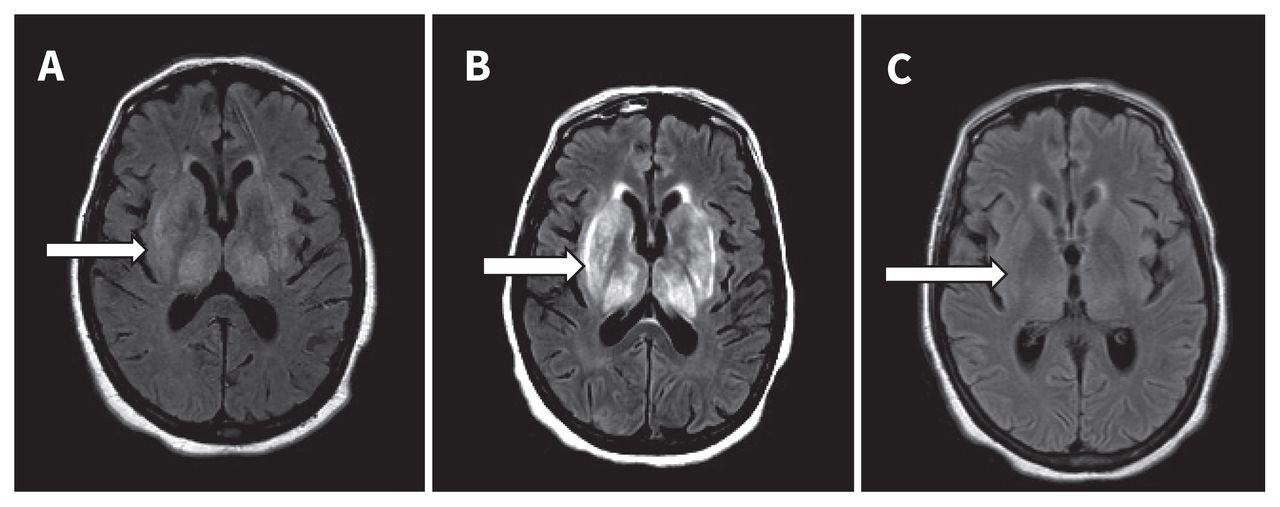
Magnetic resonance imaging (MRI) of the brain of a 68-year-old woman with autoimmune encephalitis. (A) Initial brain MRI revealed bilateral confluent hyperintense signal changes in thalami, caudate and putamen nuclei on axial fluid-attenuated inversion recovery (FLAIR) sequence (arrow). (B) Repeat brain MRI after 2 weeks showed an increase in the degree of bilateral symmetric FLAIR hyperintensity within the basal ganglia (arrow). (C) Repeat brain MRI 8 days postplasmapheresis showed nearly complete resolution of the confluent basal ganglia hyperintensity (arrow).

The patient received a 1 g dose of methylprednisolone, after which gastrointestinal bleeding developed. Two weeks later, a repeat brain MRI showed interval worsening of findings (Figure 1B). She was treated with 5 rounds of plasmapheresis, after which a repeat brain MRI showed nearly complete resolution of basal ganglia hyperintensity (Figure 1C). She received rituximab, and 6 weeks later she had made a full neurologic recovery.

Autoimmune encephalitis has an incidence of 5–10 per 100 000 person-years.1 Diagnosis is based on the clinical presentation of progressive encephalomyelitis and neuroimaging findings suggestive of encephalitis.1,2 Negative antibody test results do not exclude autoimmune encephalitis, and there is an increased risk of morbidity and death in untreated patients.2 Timely diagnosis and intervention, including immunomodulatory therapies such as intravenous immunoglobulin, high-dose corticosteroids and plasmapheresis, are associated with good outcomes. Patients with an aggressive or relapsing course require maintenance therapy with immunosuppressants.

Acknowledgement

The authors thank Stephanie Stebens for her assistance with this manuscript.

Footnotes

  • Competing interests: None declared.

  • This article has been peer reviewed.

  • The authors have obtained patient consent.

References

  1. ↵
    1. Graus F,
    2. Titulaer MJ,
    3. Balu R,
    4. et al
    . A clinical approach to the diagnosis of autoimmune encephalitis. Lancet Neurol 2016;15:391–404.
    OpenUrlCrossRefPubMed
  2. ↵
    1. Dutra LA,
    2. Abrantes F,
    3. Toso FF,
    4. et al
    . Autoimmune encephalitis: a review of diagnosis and treatment. Arq Neuropsiquiatr 2018;76:41–9.
    OpenUrlCrossRefPubMed
PreviousNext
Back to top

In this issue

Canadian Medical Association Journal: 192 (48)
CMAJ
Vol. 192, Issue 48
30 Nov 2020
  • Table of Contents
  • Index by author

Article tools

Respond to this article
Print
Download PDF
Article Alerts
To sign up for email alerts or to access your current email alerts, enter your email address below:
Email Article

Thank you for your interest in spreading the word on CMAJ.

NOTE: We only request your email address so that the person you are recommending the page to knows that you wanted them to see it, and that it is not junk mail. We do not capture any email address.

Enter multiple addresses on separate lines or separate them with commas.
Seronegative autoimmune encephalitis
(Your Name) has sent you a message from CMAJ
(Your Name) thought you would like to see the CMAJ web site.
CAPTCHA
This question is for testing whether or not you are a human visitor and to prevent automated spam submissions.
Citation Tools
Seronegative autoimmune encephalitis
Owais Alsrouji, Anza B. Memon
CMAJ Nov 2020, 192 (48) E1638; DOI: 10.1503/cmaj.200424

Citation Manager Formats

  • BibTeX
  • Bookends
  • EasyBib
  • EndNote (tagged)
  • EndNote 8 (xml)
  • Medlars
  • Mendeley
  • Papers
  • RefWorks Tagged
  • Ref Manager
  • RIS
  • Zotero
‍ Request Permissions
Share
Seronegative autoimmune encephalitis
Owais Alsrouji, Anza B. Memon
CMAJ Nov 2020, 192 (48) E1638; DOI: 10.1503/cmaj.200424
Digg logo Reddit logo Twitter logo Facebook logo Google logo Mendeley logo
  • Tweet Widget
  • Facebook Like
  • Google Plus One

Jump to section

  • Article
    • Acknowledgement
    • Footnotes
    • References
  • Figures & Tables
  • Related Content
  • Responses
  • Metrics
  • PDF

Related Articles

  • PubMed
  • Google Scholar

Cited By...

  • No citing articles found.
  • Google Scholar

More in this TOC Section

  • “Superscan” in diffusion-weighted imaging with background body suppression magnetic resonance imaging
  • Using intranasal corticosteroids
  • Langerhans cell histiocytosis in a 5-month-old baby
Show more Practice

Similar Articles

Content

  • Current issue
  • Past issues
  • Collections
  • Sections
  • Blog
  • Podcasts
  • Alerts
  • RSS
  • Early releases

Information for

  • Advertisers
  • Authors
  • Reviewers
  • CMA Members
  • Media
  • Reprint requests
  • Subscribers

About

  • General Information
  • Journal staff
  • Editorial Board
  • Governance Council
  • Journal Oversight
  • Careers
  • Contact
  • Copyright and Permissions

Copyright 2021, Joule Inc. or its licensors. All rights reserved. ISSN 1488-2329 (e) 0820-3946 (p)

All editorial matter in CMAJ represents the opinions of the authors and not necessarily those of the Canadian Medical Association or its subsidiaries.

Powered by HighWire