


Pregnant women in Canada and the United States are increasingly undergoing medical scans that pose radiation risks to their babies, researchers warn. Use of computed tomography (CT) scans during pregnancy doubled in Ontario and increased nearly four-fold in six US health systems over the past two decades, according to a large study of more than 3.4 million pregnancies published in JAMA Network Open.
“Continuing to monitor imaging rates in pregnant women is important to avoid unnecessary testing and ionizing radiation exposure to the woman and fetus,” the study authors concluded.
The study compared trends in the use of different methods of medical imaging between 1996 and 2016. Overall, 3.6% of pregnant women in Ontario and 5.3% at the US sites underwent medical imaging with ionizing radiation, mostly x-rays and CT scans. Exposure to ionizing radiation is linked to birth defects, developmental delays and cancer. Because of these risks, ultrasounds have increasingly replaced x-rays for monitoring pregnancy complications in recent decades. But the use of CT during pregnancy is still growing, as is the use of nuclear imaging during pregnancy in Ontario, even though both methods deliver far higher doses of radiation than x-rays.
The rise in CT during pregnancy mirrored use in the general population – in both groups, CT rates increased three to five times between 1996 and 2010. Patient demand and doctors practicing “defensive” medicine to avoid complaints may be contributing to these trends, the study authors suggested.
More complicated pregnancies may also require more investigation. Imaging rates were highest among women younger than 20 or older than 40, those who gave birth preterm and, in the US, among black, Native American and Hispanic women. “Minority women and younger women, compared with their counterparts, might be seen more often in emergency settings for abdominal pain where imaging is performed for clinical workup,” the study authors explained.
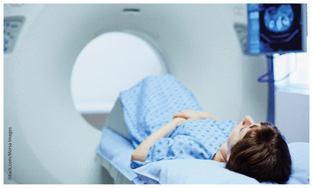
Rates of pregnant women undergoing computed tomography doubled in Ontario and increased nearly four-fold in the United States over the past two decades.
Image courtesy of iStock.com/Morsa Images
Inconsistent guidance is part of the problem, too, they noted. For example, CT is often used to diagnose pulmonary embolism, a common cause of maternal death. However, “diagnostic criteria for the management of suspected pulmonary embolism in pregnant women have largely been gleaned from studies in non-pregnant populations and retrospective studies and, thus, remain inconsistent and under debate.”
A 2015 review by the Canadian Agency for Drugs and Technology in Health found, in most instances, no statistically significant difference in outcomes in pregnant women exposed to ionizing radiation compared to those who were not exposed. However, given the limitations of the evidence, the agency recommended doctors weigh the risks and benefits and take care to minimize exposure to the fetus.
It’s not possible to tell from the JAMA Network Open study which scans were necessary or not. The authors also noted that their estimates may be conservative, because they didn’t investigate repeat examinations on the same day or imaging during pregnancies that didn’t result in a live birth.
Footnotes
Posted on cmajnews.com on Sept. 18, 2019
In this issue

Article tools


Related Articles
Cited By...
- No citing articles found.
More in this TOC Section
Similar Articles

