
Main menu

  • Home
  • Content
    • Current issue
    • Past issues
    • Early releases
    • Collections
    • Sections
    • Blog
    • Infographics & illustrations
    • Podcasts
    • COVID-19 Articles
  • Authors
    • Overview for authors
    • Submission guidelines
    • Submit a manuscript
    • Forms
    • Editorial process
    • Editorial policies
    • Peer review process
    • Publication fees
    • Reprint requests
    • Open access
  • CMA Members
    • Overview for members
    • Earn CPD Credits
    • Print copies of CMAJ
  • Subscribers
    • General information
    • View prices
  • Alerts
    • Email alerts
    • RSS
  • JAMC
    • À propos
    • Numéro en cours
    • Archives
    • Sections
    • Abonnement
    • Alertes
    • Trousse média 2022
  • CMAJ JOURNALS
    • CMAJ Open
    • CJS
    • JAMC
    • JPN

User menu

Search

  • Advanced search
CMAJ
  • CMAJ JOURNALS
    • CMAJ Open
    • CJS
    • JAMC
    • JPN
CMAJ

Advanced Search

  • Home
  • Content
    • Current issue
    • Past issues
    • Early releases
    • Collections
    • Sections
    • Blog
    • Infographics & illustrations
    • Podcasts
    • COVID-19 Articles
  • Authors
    • Overview for authors
    • Submission guidelines
    • Submit a manuscript
    • Forms
    • Editorial process
    • Editorial policies
    • Peer review process
    • Publication fees
    • Reprint requests
    • Open access
  • CMA Members
    • Overview for members
    • Earn CPD Credits
    • Print copies of CMAJ
  • Subscribers
    • General information
    • View prices
  • Alerts
    • Email alerts
    • RSS
  • JAMC
    • À propos
    • Numéro en cours
    • Archives
    • Sections
    • Abonnement
    • Alertes
    • Trousse média 2022
  • Visit CMAJ on Facebook
  • Follow CMAJ on Twitter
  • Follow CMAJ on Pinterest
  • Follow CMAJ on Youtube
  • Follow CMAJ on Instagram
Practice

Intraocular foreign bodies

Kunyong Xu and David R.P. Almeida
CMAJ January 30, 2017 189 (4) E163; DOI: https://doi.org/10.1503/cmaj.160428
Kunyong Xu
Department of Ophthalmology (Xu), Queen’s University, Kingston, Ont.; VitreoRetinal Surgery PA (Almeida), Minneapolis, Minn.
  • Find this author on Google Scholar
  • Find this author on PubMed
  • Search for this author on this site
David R.P. Almeida
Department of Ophthalmology (Xu), Queen’s University, Kingston, Ont.; VitreoRetinal Surgery PA (Almeida), Minneapolis, Minn.
  • Find this author on Google Scholar
  • Find this author on PubMed
  • Search for this author on this site
  • For correspondence: dalmeida@evolation-medical.com
  • Article
  • Figures & Tables
  • Responses
  • Metrics
  • PDF
Loading

Risk of intraocular foreign body is associated with mechanism of injury

Accurate history-taking that involves attention to the mechanism of ocular injury should be obtained. High-risk mechanisms for intraocular foreign bodies include grinding, hammering, metal shaving, machine yard work and exposure to explosives.1

Ophthalmic examination should include dilation of the pupil

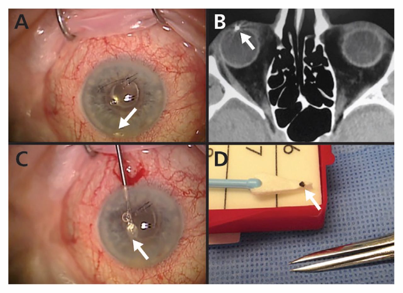
In 21%–38% of cases, intraocular foreign bodies are located in the anterior segment, allowing identification with slit-lamp examination alone.1 (Figure 1A) However, most intraocular foreign bodies are located in the posterior segment and require a dilated-pupil examination.1

Figure 1:
  • Download figure
  • Open in new tab
  • Download powerpoint
Figure 1:

Intraocular metallic foreign body embedded in the anterior chamber of the right eye. (A) Metallic foreign body in the anterior chamber (arrow). (B) Axial computed tomography scan of the orbits showing a well-defined, hyperdense foreign body producing streak artifacts in the anterior chamber (arrow). (C) Surgical removal of the foreign body (arrow). (D) The foreign body removed from the eye (arrow).

Imaging should be conducted promptly to diagnose and localize intraocular foreign bodies

Immediate computed tomography (CT) of the orbits without contrast is the modality of choice in suspected cases (Figure 1B).1 Both 1- and 3-mm cuts on CT have 100% sensitivity for detecting metallic, glass and stone intraocular foreign bodies of 0.5 mm in diameter.2 Plain-film radiography is inferior to CT, because it may miss nonmetallic intraocular foreign bodies up to 60% of the time.1

Antibiotics should be prescribed early to prevent endophthalmitis

Endophthalmitis is a complication in 3%–30% of cases involving intraocular foreign bodies.1 Therefore, patients with this injury should be given immediate treatment with prophylactic antibiotics. Systemic administration (intravenously or orally) of fluoroquinolones are good options because of their superior intraocular penetration.3

Patients with an intraocular foreign body require urgent referral to an ophthalmologist

The injured eye(s) should be protected with a rigid eye shield, and the patient should not receive anything by mouth during urgent transport to an ophthalmologist. Primary surgery to close the entry site(s) should be performed by an ophthalmologist within 12 hours after injury to reduce the risk of endophthalmitis and to prevent intraocular content extrusion.4 The foreign body must be removed; however, there is disagreement about the time frame for removal.5

Acknowledgement

The authors thank John B. Davies for providing the figures.

Footnotes

  • Competing interests: None declared.

  • This article has been peer reviewed.

References

  1. ↵
    1. Parke DW III.,
    2. Flynn HW Jr.,
    3. Fisher YL
    . Management of intraocular foreign bodies: a clinical flight plan. Can J Ophthalmol 2013;48:8–12.
    OpenUrl
  2. ↵
    1. Dass AB,
    2. Ferrone PJ,
    3. Chu YR,
    4. et al
    . Sensitivity of spiral computed tomography scanning for detecting intraocular foreign bodies. Ophthalmology 2001; 108:2326–8.
    OpenUrl
  3. ↵
    1. Hariprasad SM,
    2. Shah GK,
    3. Mieler WF,
    4. et al
    . Vitreous and aqueous penetration of orally administered moxifloxacin in humans. Arch Ophthalmol 2006; 124: 178–82.
    OpenUrlCrossRefPubMed
  4. ↵
    1. Essex RW,
    2. Yi Q,
    3. Charles PG,
    4. et al
    . Post-traumatic endopthalmitis. Ophthalmology 2004;111:2015–22.
    OpenUrlCrossRefPubMed
  5. ↵
    1. Thompson WS,
    2. Rubsamen PE,
    3. Flynn HW Jr.,
    4. et al
    . Endophthalmitis after penetrating trauma: risk factors and visual acuity outcomes. Ophthalmology 1995; 102:1696–701.
    OpenUrlPubMed
PreviousNext
Back to top

In this issue

Canadian Medical Association Journal: 189 (4)
CMAJ
Vol. 189, Issue 4
30 Jan 2017
  • Table of Contents
  • Index by author

Article tools

Respond to this article
Print
Download PDF
Article Alerts
To sign up for email alerts or to access your current email alerts, enter your email address below:
Email Article

Thank you for your interest in spreading the word on CMAJ.

NOTE: We only request your email address so that the person you are recommending the page to knows that you wanted them to see it, and that it is not junk mail. We do not capture any email address.

Enter multiple addresses on separate lines or separate them with commas.
Intraocular foreign bodies
(Your Name) has sent you a message from CMAJ
(Your Name) thought you would like to see the CMAJ web site.
CAPTCHA
This question is for testing whether or not you are a human visitor and to prevent automated spam submissions.
Citation Tools
Intraocular foreign bodies
Kunyong Xu, David R.P. Almeida
CMAJ Jan 2017, 189 (4) E163; DOI: 10.1503/cmaj.160428

Citation Manager Formats

  • BibTeX
  • Bookends
  • EasyBib
  • EndNote (tagged)
  • EndNote 8 (xml)
  • Medlars
  • Mendeley
  • Papers
  • RefWorks Tagged
  • Ref Manager
  • RIS
  • Zotero
‍ Request Permissions
Share
Intraocular foreign bodies
Kunyong Xu, David R.P. Almeida
CMAJ Jan 2017, 189 (4) E163; DOI: 10.1503/cmaj.160428
Digg logo Reddit logo Twitter logo Facebook logo Google logo Mendeley logo
  • Tweet Widget
  • Facebook Like

Jump to section

  • Article
    • Risk of intraocular foreign body is associated with mechanism of injury
    • Ophthalmic examination should include dilation of the pupil
    • Imaging should be conducted promptly to diagnose and localize intraocular foreign bodies
    • Antibiotics should be prescribed early to prevent endophthalmitis
    • Patients with an intraocular foreign body require urgent referral to an ophthalmologist
    • Acknowledgement
    • Footnotes
    • References
  • Figures & Tables
  • Responses
  • Metrics
  • PDF

Related Articles

  • No related articles found.
  • PubMed
  • Google Scholar

Cited By...

  • No citing articles found.
  • Google Scholar

More in this TOC Section

  • Pharyngeal tularemia acquired in an urban setting in Canada
  • Antithrombotic agents in patients with COVID-19
  • Bullous pemphigoid associated with use of dipeptidyl peptidase-4 inhibitor
Show more Practice

Similar Articles

Collections

  • Sections
    • Five Things to Know About
  • Topics
    • Emergency medicine
    • Ophthalmology
    • Trauma, injury & trauma surgery


View Latest Classified Ads

Content

  • Current issue
  • Past issues
  • Collections
  • Sections
  • Blog
  • Podcasts
  • Alerts
  • RSS
  • Early releases

Information for

  • Advertisers
  • Authors
  • Reviewers
  • CMA Members
  • Media
  • Reprint requests
  • Subscribers

About

  • General Information
  • Journal staff
  • Editorial Board
  • Advisory Panels
  • Governance Council
  • Journal Oversight
  • Careers
  • Contact
  • Copyright and Permissions
  • Accessibiity
  • CMA Civility Standards
CMAJ Group

Copyright 2022, CMA Impact Inc. or its licensors. All rights reserved. ISSN 1488-2329 (e) 0820-3946 (p)

All editorial matter in CMAJ represents the opinions of the authors and not necessarily those of the Canadian Medical Association or its subsidiaries.

To receive any of these resources in an accessible format, please contact us at CMAJ Group, 500-1410 Blair Towers Place, Ottawa ON, K1J 9B9; p: 1-888-855-2555; e: cmajgroup@cmaj.ca

Powered by HighWire