
Main menu

  • Home
  • Content
    • Current issue
    • Past issues
    • Early releases
    • Collections
    • Sections
    • Blog
    • Infographics & illustrations
    • Podcasts
    • COVID-19 Articles
    • Obituary notices
  • Authors & Reviewers
    • Overview for authors
    • Submission guidelines
    • Submit a manuscript
    • Forms
    • Editorial process
    • Editorial policies
    • Peer review process
    • Publication fees
    • Reprint requests
    • Open access
    • Patient engagement
  • Members & Subscribers
    • Benefits for CMA Members
    • CPD Credits for Members
    • Subscribe to CMAJ Print
    • Subscription Prices
    • Obituary notices
  • Alerts
    • Email alerts
    • RSS
  • JAMC
    • À propos
    • Numéro en cours
    • Archives
    • Sections
    • Abonnement
    • Alertes
    • Trousse média 2023
    • Avis de décès
  • CMAJ JOURNALS
    • CMAJ Open
    • CJS
    • JAMC
    • JPN

User menu

Search

  • Advanced search
CMAJ
  • CMAJ JOURNALS
    • CMAJ Open
    • CJS
    • JAMC
    • JPN
CMAJ

Advanced Search

  • Home
  • Content
    • Current issue
    • Past issues
    • Early releases
    • Collections
    • Sections
    • Blog
    • Infographics & illustrations
    • Podcasts
    • COVID-19 Articles
    • Obituary notices
  • Authors & Reviewers
    • Overview for authors
    • Submission guidelines
    • Submit a manuscript
    • Forms
    • Editorial process
    • Editorial policies
    • Peer review process
    • Publication fees
    • Reprint requests
    • Open access
    • Patient engagement
  • Members & Subscribers
    • Benefits for CMA Members
    • CPD Credits for Members
    • Subscribe to CMAJ Print
    • Subscription Prices
    • Obituary notices
  • Alerts
    • Email alerts
    • RSS
  • JAMC
    • À propos
    • Numéro en cours
    • Archives
    • Sections
    • Abonnement
    • Alertes
    • Trousse média 2023
    • Avis de décès
  • Visit CMAJ on Facebook
  • Follow CMAJ on Twitter
  • Follow CMAJ on Pinterest
  • Follow CMAJ on Youtube
  • Follow CMAJ on Instagram
Practice

Accidental barium bronchography

Ahmad Zaheen and Matthew B. Stanbrook
CMAJ May 08, 2017 189 (18) E670; DOI: https://doi.org/10.1503/cmaj.160969
Ahmad Zaheen
Department of Medicine (Zaheen), University of Toronto; Division of Respirology (Stanbrook), Toronto Western Hospital, Toronto, Ont.
  • Find this author on Google Scholar
  • Find this author on PubMed
  • Search for this author on this site
Matthew B. Stanbrook
Department of Medicine (Zaheen), University of Toronto; Division of Respirology (Stanbrook), Toronto Western Hospital, Toronto, Ont.
  • Find this author on Google Scholar
  • Find this author on PubMed
  • Search for this author on this site
  • For correspondence: matthew.stanbrook@uhn.ca
  • Article
  • Figures & Tables
  • Responses
  • Metrics
  • PDF
Loading

A 46-year-old man with a history of T2N2 right tonsillar squamous cell carcinoma was referred to the emergency department after a large aspiration event during a barium esophogram. His cancer had been treated five years previously with panitumumab, radiation and salvage surgery for residual tumour. The esophogram was arranged for a six-month history of dysphagia and periprandial coughing up of food contents. The patient was asymptomatic upon arrival in the emergency department.

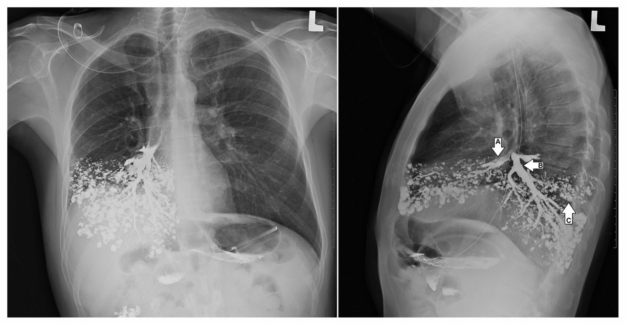
Upon examination, he appeared well and hemodynamically stable, with a respiratory rate of 20 breaths per minute, oxygen saturation of 94% on ambient air and a temperature of 38.1°C. Respiratory examination showed no increased work of breathing, with crackles in the right lung base on auscultation. Neurologic deficits included palsy of the right hypoglossal nerve and asymmetry of the soft palate. Chest radiographs conducted in the emergency department showed accumulation of barium in the right middle and lower lobes (Figure 1). Bronchoscopy confirmed localization of barium in the right lung, with no evidence of tracheoesophageal fistula. Nasopharyngoscopy showed normal vocal cord function, but absent supraglottic and glottic sensation. A videofluoroscopic swallowing examination identified uncoordinated swallowing with florid aspiration, and a diagnosis of aspiration pneumonitis was made. These findings were attributed to delayed effects from radiation treatment. He remained clinically stable and a feeding gastrostomy tube was inserted.

Figure 1:
  • Download figure
  • Open in new tab
  • Download powerpoint
Figure 1:

Radiographs of the posteroanterior (left image) and lateral (right image) chest showing barium bronchograms of the right middle (arrow A) and right lower (arrow B) lobe bronchi. Extensive tree-in-bud opacifications in the right middle and lower lobes are indicative of bronchiolar filling (arrow C).

Risk factors for aspiration include altered level of consciousness, neurologic dysfunction of the upper airway, structural esophageal disease, cancers of the aerodigestive tract and iatrogenic disruption of the airway (i.e., intubation and endoscopy).1 The anatomic configuration of the bronchial tree predisposes contents to enter the right and middle lobes on erect aspiration, as shown in our case.

Aspiration pneumonitis is an inflammatory injury to the lung triggered by exposure to noxious stimuli, typically of gastric origin. Clinical manifestations range from none to cough, wheeze, dyspnea, respiratory failure and, in severe cases, acute respiratory distress syndrome.1 Treatment is typically supportive, and routine antibiotic use is not indicated.1

Barium is chemically inert and had been used as a contrast medium for bronchography before the advent of computed tomography and bronchoscopy.2 Although historically found to be safe in this context, aspiration of barium may also produce a chemical pneumonitis in some cases.3

Footnotes

  • Competing interests: None declared.

  • This article has been peer reviewed.

  • The authors have obtained patient consent.

  • Disclaimer: Matthew Stanbrook is a deputy editor for CMAJ and was not involved in the editorial decision-making process for this article.

References

  1. ↵
    1. Marik PE
    . Aspiration pneumonitis and aspiration pneumonia. N Engl J Med 2001;344:665–71.
    OpenUrlCrossRefPubMed
  2. ↵
    1. Shook CD,
    2. Felson B
    . Inhalation bronchography. Chest 1970;58:333–7.
    OpenUrlCrossRefPubMed
  3. ↵
    1. Hundemer GL,
    2. Kumar V,
    3. Vaduganathan M
    . Large-volume barium aspiration. Proc Bayl Univ Med Cent 2015;28:183–4.
    OpenUrl
PreviousNext
Back to top

In this issue

Canadian Medical Association Journal: 189 (18)
CMAJ
Vol. 189, Issue 18
8 May 2017
  • Table of Contents
  • Index by author

Article tools

Respond to this article
Print
Download PDF
Article Alerts
To sign up for email alerts or to access your current email alerts, enter your email address below:
Email Article

Thank you for your interest in spreading the word on CMAJ.

NOTE: We only request your email address so that the person you are recommending the page to knows that you wanted them to see it, and that it is not junk mail. We do not capture any email address.

Enter multiple addresses on separate lines or separate them with commas.
Accidental barium bronchography
(Your Name) has sent you a message from CMAJ
(Your Name) thought you would like to see the CMAJ web site.
CAPTCHA
This question is for testing whether or not you are a human visitor and to prevent automated spam submissions.
Citation Tools
Accidental barium bronchography
Ahmad Zaheen, Matthew B. Stanbrook
CMAJ May 2017, 189 (18) E670; DOI: 10.1503/cmaj.160969

Citation Manager Formats

  • BibTeX
  • Bookends
  • EasyBib
  • EndNote (tagged)
  • EndNote 8 (xml)
  • Medlars
  • Mendeley
  • Papers
  • RefWorks Tagged
  • Ref Manager
  • RIS
  • Zotero
‍ Request Permissions
Share
Accidental barium bronchography
Ahmad Zaheen, Matthew B. Stanbrook
CMAJ May 2017, 189 (18) E670; DOI: 10.1503/cmaj.160969
Digg logo Reddit logo Twitter logo Facebook logo Google logo Mendeley logo
  • Tweet Widget
  • Facebook Like

Jump to section

  • Article
    • Footnotes
    • References
  • Figures & Tables
  • Responses
  • Metrics
  • PDF

Related Articles

  • No related articles found.
  • PubMed
  • Google Scholar

Cited By...

  • No citing articles found.
  • Google Scholar

More in this TOC Section

  • Screening for primary aldosteronism in primary care
  • Duodenal duplication cyst in a 61-year-old man
  • A young woman with fever and polyserositis caused by familial Mediterranean fever
Show more Practice

Similar Articles


View Latest Classified Ads

Content

  • Current issue
  • Past issues
  • Collections
  • Sections
  • Blog
  • Podcasts
  • Alerts
  • RSS
  • Early releases

Information for

  • Advertisers
  • Authors
  • Reviewers
  • CMA Members
  • CPD credits
  • Media
  • Reprint requests
  • Subscribers

About

  • General Information
  • Journal staff
  • Editorial Board
  • Advisory Panels
  • Governance Council
  • Journal Oversight
  • Careers
  • Contact
  • Copyright and Permissions
  • Accessibiity
  • CMA Civility Standards
CMAJ Group

Copyright 2023, CMA Impact Inc. or its licensors. All rights reserved. ISSN 1488-2329 (e) 0820-3946 (p)

All editorial matter in CMAJ represents the opinions of the authors and not necessarily those of the Canadian Medical Association or its subsidiaries.

To receive any of these resources in an accessible format, please contact us at CMAJ Group, 500-1410 Blair Towers Place, Ottawa ON, K1J 9B9; p: 1-888-855-2555; e: cmajgroup@cmaj.ca

Powered by HighWire