


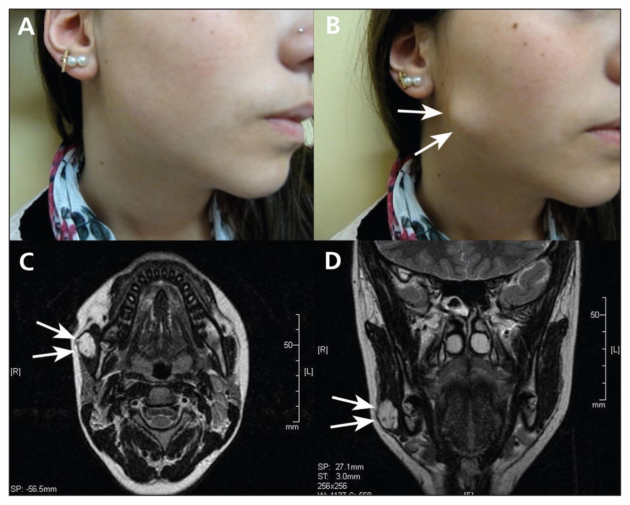
A 16-year-old girl presented with a one-year history of a mass in her right cheek that became apparent only with teeth-clenching (Figure 1A and B; video available at www.cmaj.ca/lookup/suppl/doi:10.1503/cmaj.140364/-/DC1). The lesion was soft, well-defined and painless. It measured 3 × 2 cm, and the overlying skin was normal. There was no evidence of facial nerve involvement. Examination of the oral cavity was unremarkable. Magnetic resonance imaging showed a lobulated soft tissue lesion within the right masseter (Figure 1C and D). Fine-needle aspiration cytology yielded a hemorrhagic specimen.

Cheek of a 16-year-old girl (A) at rest and (B) with a mass apparent only with teeth-clenching. (C) Axial T2-weighted magnetic resonance imaging (MRI) scan showing a hyperintense, lobulated soft tissue lesion within the right masseter. (D) Coronal MRI scan showing the lesion.
The differential diagnosis of a mass within the masseter includes congenital cyst, lymphadenopathy, sialocele of parotid duct, parotid neoplasm, lymphangioma, benign or malignant muscle tumour, masseteric hypertrophy and schwannoma. The clinical and imaging findings in this case were consistent with an intramuscular hemangioma. We recommended resection of the mass through an extraoral approach; however, the patient’s parents did not consent to the proposed treatment because of the risk of facial paralysis during the surgical procedure. The patient was lost to follow-up.
Intramuscular hemangioma represents less than 1% of all hemangiomas.1 It affects mainly the trunk and the extremities, where the muscle volume is larger. About 13% of the lesions present in the head and neck region.2 In the head, the masseter muscle is the most commonly involved site. Because of their deep location, intramuscular hemangiomas are difficult to diagnose.
The turkey wattle sign is an unusual pathognomonic manifestation of intramasseter and intraparotid hemangiomas. It refers to enlargement of the lesion with teeth-clenching or with dependent head positioning.3,4 The sign may be due to vascular engorgement within the lesion, which impedes venous return from the head to the superior vena cava. The turkey wattle is a red vascular structure in the neck of the male turkey that can increase in size when filled with blood.5
See the following video online: The patient’s cheek with a mass apparent with teeth-clenching. www.cmaj.ca/lookup/suppl/doi:10.1503/cmaj.140364/-/DC1
Footnotes
Competing interests: None declared.
This article has been peer reviewed.
The authors have obtained patient consent.
In this issue

Article tools


Related Articles
Cited By...
- No citing articles found.
More in this TOC Section
Similar Articles
Collections

