


In a recent Practice article,1 the magnetic resonance imaging scan showing mineral deposits within the globus pallidus was incorrect. The correct image (Figure 1) and revised caption are included here.
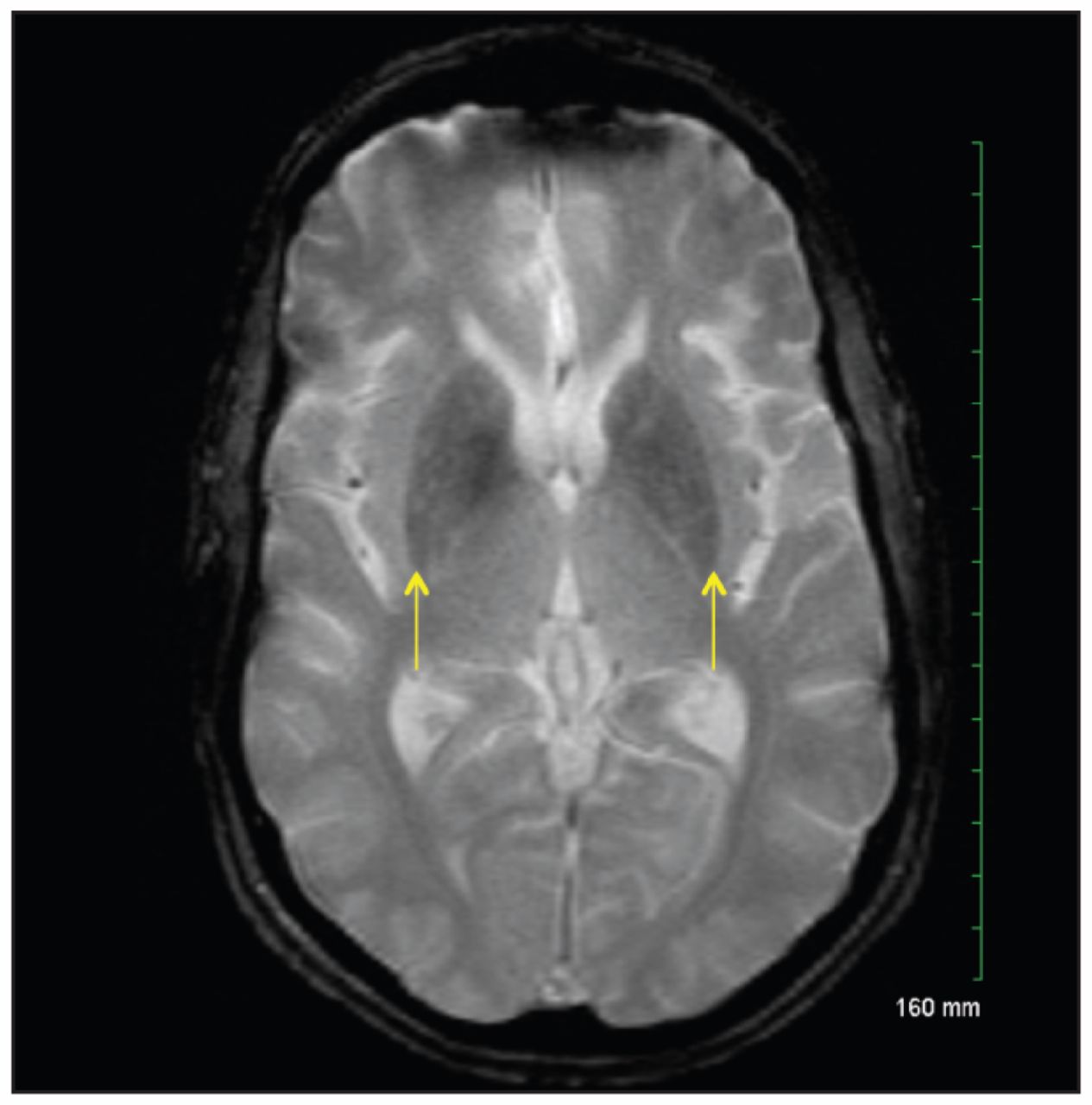
Figure 1:
Gradient-echo magnetic resonance imaging scan of the brain in case 1, showing diffuse, somewhat increased mineralization seen as low signal intensity in the basal ganglia with bilateral involvement of the putamen (arrows) and caudate nucleus. Even lower signal intensity of the medial globus pallidus is a physiological finding.
Reference
In this issue
Article tools
Respond to this article


Related Articles
Cited By...
- No citing articles found.
More in this TOC Section
Similar Articles

