
Main menu

  • Home
  • COVID-19
    • Articles & podcasts
    • Blog posts
    • Collection
    • News
  • Content
    • Current issue
    • Past issues
    • Early releases
    • Collections
    • Sections
    • Blog
    • Infographics & illustrations
    • Podcasts
    • Classified ads
  • Authors
    • Overview for authors
    • Submission guidelines
    • Submit a manuscript
    • Forms
    • Editorial process
    • Editorial policies
    • Peer review process
    • Publication fees
    • Reprint requests
  • CMA Members
    • Overview for members
    • Earn CPD Credits
    • Print copies of CMAJ
    • Career Ad Discount
  • Subscribers
    • General information
    • View prices
  • Alerts
    • Email alerts
    • RSS
  • JAMC
    • À propos
    • Numéro en cours
    • Archives
    • Sections
    • Abonnement
    • Alertes
  • CMAJ JOURNALS
    • CMAJ Open
    • CJS
    • JAMC
    • JPN

User menu

Search

  • Advanced search
CMAJ
  • CMAJ JOURNALS
    • CMAJ Open
    • CJS
    • JAMC
    • JPN
CMAJ

Advanced Search

  • Home
  • COVID-19
    • Articles & podcasts
    • Blog posts
    • Collection
    • News
  • Content
    • Current issue
    • Past issues
    • Early releases
    • Collections
    • Sections
    • Blog
    • Infographics & illustrations
    • Podcasts
    • Classified ads
  • Authors
    • Overview for authors
    • Submission guidelines
    • Submit a manuscript
    • Forms
    • Editorial process
    • Editorial policies
    • Peer review process
    • Publication fees
    • Reprint requests
  • CMA Members
    • Overview for members
    • Earn CPD Credits
    • Print copies of CMAJ
    • Career Ad Discount
  • Subscribers
    • General information
    • View prices
  • Alerts
    • Email alerts
    • RSS
  • JAMC
    • À propos
    • Numéro en cours
    • Archives
    • Sections
    • Abonnement
    • Alertes
  • Visit CMAJ on Facebook
  • Follow CMAJ on Twitter
  • Follow CMAJ on Pinterest
  • Follow CMAJ on Youtube
  • Follow CMAJ on Instagram
Practice

Portal venous gas and pneumatosis intestinalis

Shung-Wei Liu and Tai-Yu Huang
CMAJ February 19, 2013 185 (3) E175; DOI: https://doi.org/10.1503/cmaj.120219
Shung-Wei Liu
From the Department of Emergency Medicine, Buddhist Tzu Chi General Hospital, Hualien, Taiwan
  • Find this author on Google Scholar
  • Find this author on PubMed
  • Search for this author on this site
Tai-Yu Huang
From the Department of Emergency Medicine, Buddhist Tzu Chi General Hospital, Hualien, Taiwan
  • Find this author on Google Scholar
  • Find this author on PubMed
  • Search for this author on this site
  • For correspondence: huangtaiyu72@gmail.com
  • Article
  • Figures & Tables
  • Related Content
  • Responses
  • Metrics
  • PDF
Loading

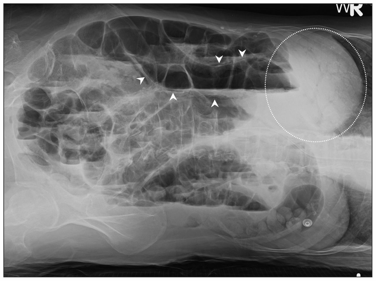
A 60-year-old woman with a history of atrial fibrillation presented after one day of drowsiness and abdominal bloating. Examination revealed fever, a distended abdomen without muscle guarding and decreased bowel sounds. Results of blood tests and computed tomography (CT) scans of the head showed no specific abnormalities. A radiograph taken with the patient in the left lateral decubitus position to rule out acute abdomen showed branching radiolucency in the liver and gas in the bowel wall (Figure 1). Subsequent contrast-enhanced CT scans of the abdomen revealed a left atrial thrombus, portal venous gas, a filling defect in the superior mesenteric artery and pneumatosis intestinalis (Appendix 1, available at www.cmaj.ca/lookup/suppl/doi:10.1503/cmaj.120219/-/DC1). Acute mesenteric infarction secondary to thromboembolism of cardiac origin was diagnosed. Because of septic shock, the patient died on the operating table before emergency surgery was started.

Figure 1:
  • Download figure
  • Open in new tab
  • Download powerpoint
Figure 1:

Radiograph of a 60-year-old woman with a distended abdomen taken in the left lateral decubitus position, showing branching air in the liver (circle) and gas in the bowel wall (arrowheads).

Portal venous gas and pneumatosis intestinalis, first described in 1955 and 1963 respectively, are radiographic signs that have been traditionally associated with bowel ischemia.1 Radiographic criteria for portal venous gas include a branching radiolucency that extends to within 2 cm of the liver capsule; this finding must be differentiated from that of pneumobilia (gas in the biliary system), which is visualized as air located centrally within the liver.1 Pneumatosis intestinalis is defined as the presence of gas within the bowel wall and is characterized by subserosal or submucosal gas-filled cysts in the gastrointestinal tract. Because of the widespread use of CT imaging, many nonischemic causes have been recently reported, including infectious, neoplastic and inflammatory bowel diseases for pneumatosis intestinalis, and portal pylephlebitis, abdominal trauma and iatrogenic procedures for portal venous gas.1,2 Therefore, neither of the radiographic signs of portal venous gas or pneumatosis intestinalis alone is definitive for diagnosing bowel ischemia. However, the simultaneous presence of both findings is more predictive of transmural bowel ischemia and higher mortality.1 If mesenteric ischemia is suspected, CT scanning is considered first-line imaging.2,3 However, appropriate interpretation of subtle findings on plain radiographs may help to make an early diagnosis of bowel ischemia.2,3

Footnotes

  • Competing interests: None declared.

  • This article has been peer reviewed.

References

  1. ↵
    1. Wiesner W,
    2. Mortelé KJ,
    3. Glickman JN,
    4. et al
    . Pneumatosis intestinalis and portomesenteric venous gas in intestinal ischemia: correlation of CT findings with severity of ischemia and clinical outcome. AJR Am J Roentgenol 2001;177:1319–23.
    OpenUrlPubMed
  2. ↵
    1. Wayne E,
    2. Ough M,
    3. Wu A,
    4. et al
    . Management algorithm for pneumatosis intestinalis and portal venous gas: treatment and outcome of 88 consecutive cases. J Gastrointest Surg 2010;14:437–48.
    OpenUrlCrossRefPubMed
  3. ↵
    1. Menke J
    . Diagnostic accuracy of multidetector CT in acute mesenteric ischemia: systematic review and meta-analysis. Radiology 2010;256:93–101.
    OpenUrlCrossRefPubMed
PreviousNext
Back to top

In this issue

Canadian Medical Association Journal: 185 (3)
CMAJ
Vol. 185, Issue 3
19 Feb 2013
  • Table of Contents
  • Index by author

Article tools

Respond to this article
Print
Download PDF
Article Alerts
To sign up for email alerts or to access your current email alerts, enter your email address below:
Email Article

Thank you for your interest in spreading the word on CMAJ.

NOTE: We only request your email address so that the person you are recommending the page to knows that you wanted them to see it, and that it is not junk mail. We do not capture any email address.

Enter multiple addresses on separate lines or separate them with commas.
Portal venous gas and pneumatosis intestinalis
(Your Name) has sent you a message from CMAJ
(Your Name) thought you would like to see the CMAJ web site.
CAPTCHA
This question is for testing whether or not you are a human visitor and to prevent automated spam submissions.
Citation Tools
Portal venous gas and pneumatosis intestinalis
Shung-Wei Liu, Tai-Yu Huang
CMAJ Feb 2013, 185 (3) E175; DOI: 10.1503/cmaj.120219

Citation Manager Formats

  • BibTeX
  • Bookends
  • EasyBib
  • EndNote (tagged)
  • EndNote 8 (xml)
  • Medlars
  • Mendeley
  • Papers
  • RefWorks Tagged
  • Ref Manager
  • RIS
  • Zotero
‍ Request Permissions
Share
Portal venous gas and pneumatosis intestinalis
Shung-Wei Liu, Tai-Yu Huang
CMAJ Feb 2013, 185 (3) E175; DOI: 10.1503/cmaj.120219
Digg logo Reddit logo Twitter logo Facebook logo Google logo Mendeley logo
  • Tweet Widget
  • Facebook Like
  • Google Plus One

Jump to section

  • Article
    • Footnotes
    • References
  • Figures & Tables
  • Related Content
  • Responses
  • Metrics
  • PDF

Related Articles

  • PubMed
  • Google Scholar

Cited By...

  • No citing articles found.
  • Google Scholar

More in this TOC Section

  • Azathioprine-induced severe anemia potentiated by the concurrent use of allopurinol
  • Schwannoma of the tongue
  • “Superscan” in diffusion-weighted imaging with background body suppression magnetic resonance imaging
Show more Practice

Similar Articles

Collections

  • Sections
    • Clinical Images
  • Topics
    • Imaging
    • Gastroenterology

Content

  • Current issue
  • Past issues
  • Collections
  • Sections
  • Blog
  • Podcasts
  • Alerts
  • RSS
  • Early releases

Information for

  • Advertisers
  • Authors
  • Reviewers
  • CMA Members
  • Media
  • Reprint requests
  • Subscribers

About

  • General Information
  • Journal staff
  • Editorial Board
  • Governance Council
  • Journal Oversight
  • Careers
  • Contact
  • Copyright and Permissions

Copyright 2021, Joule Inc. or its licensors. All rights reserved. ISSN 1488-2329 (e) 0820-3946 (p)

All editorial matter in CMAJ represents the opinions of the authors and not necessarily those of the Canadian Medical Association or its subsidiaries.

Powered by HighWire