


Article Figures & Tables
Figures
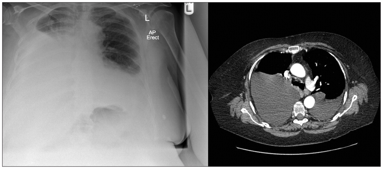
Figure 1: Chest radiograph and contrast-enhanced computed tomography scan of the thorax showing bilateral pleural effusion in a 50-year-old woman with diffuse large B-cell lymphoma.
Figure 2: Milky off-white to yellow pleural aspirate.
Figure 3: The pathway of the thoracic duct. Reprinted with permission. 1 Copyright © 1992 American College of Chest Physicians.
Box 1: Differential diagnosis of bilateral pleural effusion 4
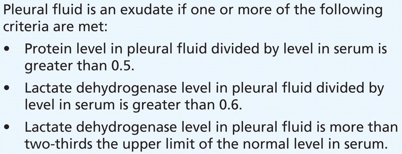
Box 2: Light’s criteria for classification of an effusion 5


Tables
Table 1: Routine and additional analyses performed on pleural fluid (4)


In this issue
Article tools
Respond to this article


Jump to section
Related Articles
Cited By...
- No citing articles found.
More in this TOC Section
Similar Articles
Collections

