


Article Figures & Tables
Figures
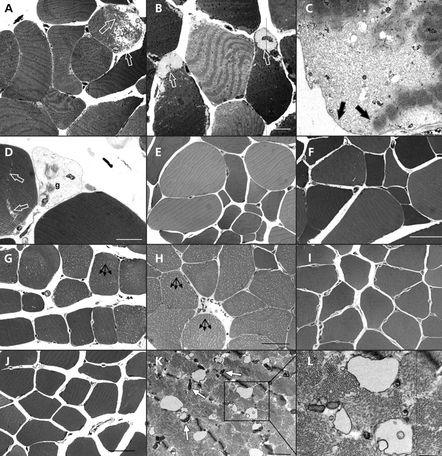
Figure 1: Skeletal muscle injury in patients with statin-associated myopathy. Light micrographs (A, B, D, E, F) and an electron micrograph (C) of transverse sections through the vastus lateralis muscle of patients with myopathy and current (B, D, E, F) or previous (A, C) statin use. Subsarcolemmal detachment of myofibrils is indicated by arrows in (A), (B) and (C). In (D), a ghost cell (g) is seen next to a developing split-fibre (arrows). (G, H): Light micrographs of semi-thin, transverse sections through the vastus lateralis muscle of myopathic patients currently (G) or formerly taking statins (H). Vacuolization is seen in most of the fibres (arrows). (I, J): The muscles fibres from control patients show no vacuolization. (K, L): Electron micrographs of a transverse section through the vastus lateralis muscle from a patient with myopathy who formerly used statins; intracellular vacuoles are present. The boxed area in (K) is enlarged in (L). The T-tubules are grossly vacuolated, but the adjacent mitochondria (m) have a normal appearance. Bars: A, B = 10 μm; C = 0.2 μm; D = 3 μm; E, F = 50 μm; G, I, J = 50 μm, H = 30 μm, K = 1 μm; L = 0.5 μm.
Figure 2: Fluorescence micrographs of transverse ultra-thin frozen sections labelled with a T-tubular marker (annexin A6, green) and a nuclear marker (Hoechst, blue) showing vacuolization (arrows) in a patient with statin-associated myopathy (A). The T-tubular system is undamaged in a control patient (B). Bar = 10 μm.
Figure 3: The levels of mRNA expression of 8 selected genes that code for components of calcium release or uptake. The expression of each gene was normalized to the amount of 18S RNA in the sample. Shown are the mean values with the standard error of the mean. Patients were grouped by the presence of significant muscle damage (squares, n = 25) or no significant damage (circles, n = 32). Note: RYR1 = ryanodine receptor 1, RYR3 = ryanodine receptor 3, ITPR1 = inositol 1,4,5-triphosphate receptor, type 1, ITPR2 = inositol 1,4,5-triphosphate receptor, type 2, ITPR3 = inositol 1,4,5-triphosphate receptor, type 3; SERCA1 = sarco-endoplasmic reticulum transporting Ca2+ ATPase 1; SERCA2 = sarco-endoplasmic reticulum transporting Ca2+ ATPase 2; SERCA3 = sarco-endoplasmic reticulum transporting Ca2+ ATPase 3.
Figure 4: Circulating levels of creatine phosphokinase in patients with statin-associated myopathy and current (myopathy current statin) or past (myopathy former statin) statin therapy and in age-matched patients currently using statins with no myopathy. Note: ULN = upper limit of normal.


Tables
Table 1: Characteristics of patients included in a study of the relation between statin-associated myopathy and structural muscle damage

Table 2: History of myopathy among patients who used statins


In this issue
Article tools


Jump to section
Related Articles
Cited By...
- Increased remodeling and impaired adaption to endurance exercise in desminopathy
- Etanercept-induced myositis: do we have to stop it? A surprising outcome
- Authors' reply to Huffman and colleagues
- Comparison of efficacy and adverse effect profile of high dose versus standard dose atorvastatin in acute ST elevation myocardial infarction patients
More in this TOC Section
Similar Articles

