
Main menu

  • Home
  • Content
    • Current issue
    • Past issues
    • Early releases
    • Collections
    • Sections
    • Blog
    • Infographics & illustrations
    • Podcasts
    • COVID-19 Articles
    • Obituary notices
  • Authors & Reviewers
    • Overview for authors
    • Submission guidelines
    • Submit a manuscript
    • Forms
    • Editorial process
    • Editorial policies
    • Peer review process
    • Publication fees
    • Reprint requests
    • Open access
    • Patient engagement
  • Members & Subscribers
    • Benefits for CMA Members
    • CPD Credits for Members
    • Subscribe to CMAJ Print
    • Subscription Prices
    • Obituary notices
  • Alerts
    • Email alerts
    • RSS
  • JAMC
    • À propos
    • Numéro en cours
    • Archives
    • Sections
    • Abonnement
    • Alertes
    • Trousse média 2023
    • Avis de décès
  • CMAJ JOURNALS
    • CMAJ Open
    • CJS
    • JAMC
    • JPN

User menu

Search

  • Advanced search
CMAJ
  • CMAJ JOURNALS
    • CMAJ Open
    • CJS
    • JAMC
    • JPN
CMAJ

Advanced Search

  • Home
  • Content
    • Current issue
    • Past issues
    • Early releases
    • Collections
    • Sections
    • Blog
    • Infographics & illustrations
    • Podcasts
    • COVID-19 Articles
    • Obituary notices
  • Authors & Reviewers
    • Overview for authors
    • Submission guidelines
    • Submit a manuscript
    • Forms
    • Editorial process
    • Editorial policies
    • Peer review process
    • Publication fees
    • Reprint requests
    • Open access
    • Patient engagement
  • Members & Subscribers
    • Benefits for CMA Members
    • CPD Credits for Members
    • Subscribe to CMAJ Print
    • Subscription Prices
    • Obituary notices
  • Alerts
    • Email alerts
    • RSS
  • JAMC
    • À propos
    • Numéro en cours
    • Archives
    • Sections
    • Abonnement
    • Alertes
    • Trousse média 2023
    • Avis de décès
  • Visit CMAJ on Facebook
  • Follow CMAJ on Twitter
  • Follow CMAJ on Pinterest
  • Follow CMAJ on Youtube
  • Follow CMAJ on Instagram
News

Radiologists virtually unwrap mummy’s secrets

Roger Collier
CMAJ April 28, 2009 180 (9) E23-E24; DOI: https://doi.org/10.1503/cmaj.090237
Roger Collier
  • Find this author on Google Scholar
  • Find this author on PubMed
  • Search for this author on this site
  • Article
  • Figures & Tables
  • Responses
  • Metrics
  • PDF
Loading

In 1997, a radiologist in Montréal, Quebec examined a woman and found that her left femur had once been broken. That finding is now under suspicion. The radiologist said the woman’s bad teeth stemmed from too much beer. That also appears to be wrong. A recent computed tomography (CT) scan has revealed much about the health of this mysterious woman —and she only waited 2500 years for the exam. Give or take a century.

On the evening of Nov. 18, 2008, radiologists at the University of Ottawa Heart Institute slid Hetep-Bastet into a scanner’s narrow core. Accustomed to tombs, Hetep-Bastet, an Egyptian mummy dating to sometime before 500 BC, didn’t seem to mind. Dr. Andrew Nelson, the University of Western On-tario anthropologist interpreting the results, says the radiological study of mummies teaches us about the history of diseases and the general health of past cultures.

Not that academic casket-cracking is new. Researchers have been gleaning health information from linen-wrapped remains for more than a century. The Museum of Natural History, in Austria, x-rayed an Egyptian coffin (W. König 14 Photographien mit Röntgen-Strahlen, aufgenommen im Physikalis-chen Verein, Frankfurt a M. Leipzig: JA Barth, 1896) only a year after physicist Wilhelm Röntgen had taken the world’s first x-ray. Many researchers have since studied mummies, often through autopsy. That was a problem, however, because autopsies made a mess of millennium-old remains.

“The real emphasis now is to do things non-destructively,” says Nelson. “The CT scanner is ideal for that.”

Technology no doubt makes investigations safer for King Tut and cohorts, but mummy-scanning offers new challenges for researchers. A CT scan of a living person is easy to interpret because bone is much denser than live tissue. Bones hungrily absorb radiation. Muscles, fat and organs are x-ray thoroughfares. But the thoroughly dehydrated skin and flesh of a mummy is similar in density to bone. Even the wrappings eventually come to resemble fibulas more than fabric. It is also impossible to inject dye into a mummy’s bloodstream to highlight blood vessels, because mummies have no blood to stream.

Figure
  • Download figure
  • Open in new tab
  • Download powerpoint

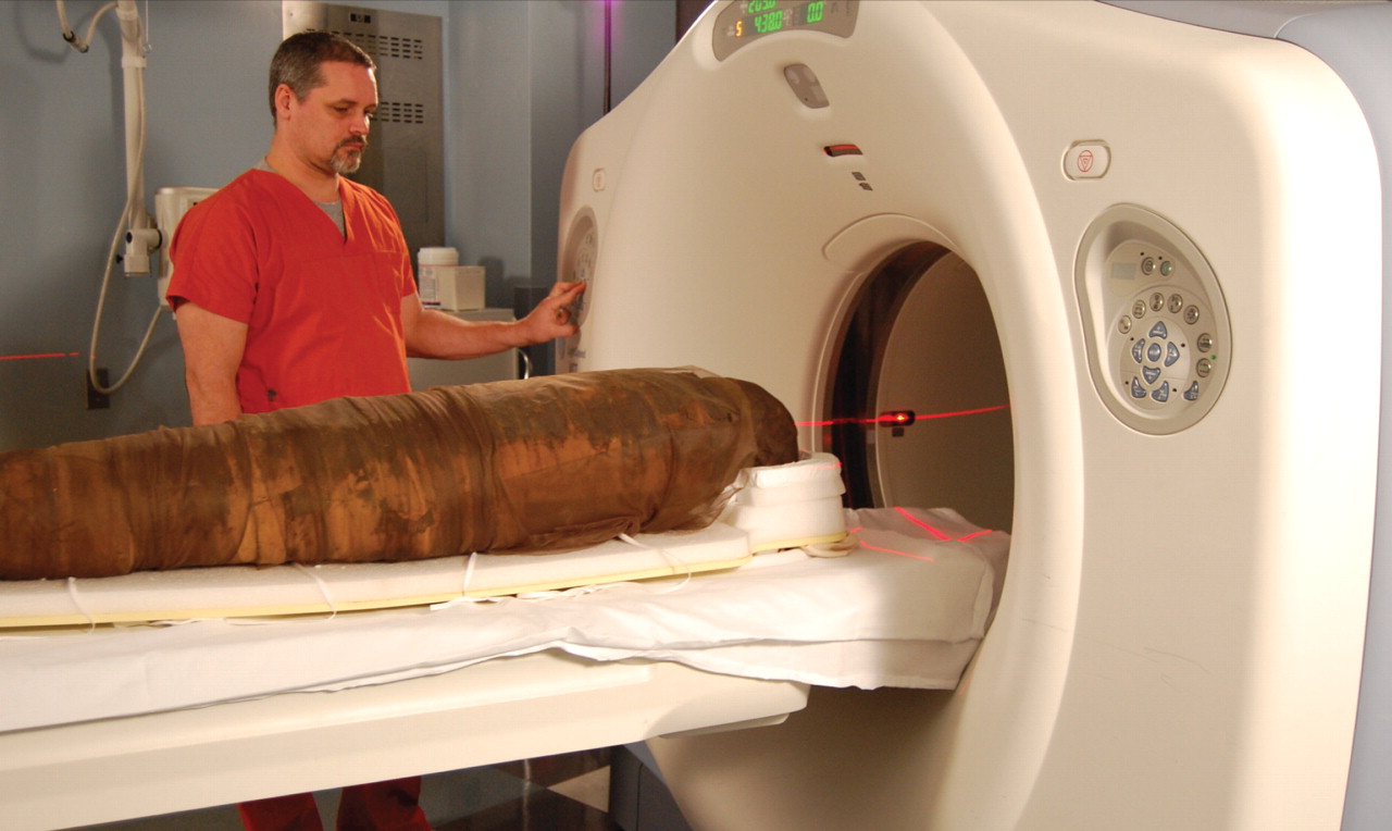
FIGURE. A computed tomography scan of Hetep-Bastet, an Egyptian mummy, was performed at The University of Ottawa Heart Institute on Nov. 18, 2008. Radiologists donated their time, and the scan took place after regular hours as to not disturb patient scans. Scanning is a nondestructive way to study ancient remains. Image courtesy of The University of Ottawa Heart Institute

Conducting the scan is a breeze. Technologists scanning a living patient must compensate for movement due to breathing, fidgeting and a beating heart. But mummies tend to remain still. They also don’t mind being bathed in radiation, so they remain in the scanner longer, allowing for better images. Capturing the data is easy. Creating an image from that data? Not so easy.

“There’s only a few seconds to several minutes of reconstruction for a living patient,” says Dr. Blair Macdonald, a staff radiologist with The Ottawa Hospital. “For a mummy, it could be in the range of hours to days to strip away various resins and identify what is soft tissue and bone.”

Fortunately, a new way of conducting CT scans is simplifying that process. The dual-energy technique, used in the Hetep-Bastet exam, involves scanning at different power levels. This allows radiologists to capture more detailed information and better discriminate between bone and soft tissue. Since all parts of mummy remains are alike in density, they are ideal for testing the technique. Radiologists can then apply the lessons learned to living patients.

“The thing that is most exciting for me is the dual-energy scanning,” says Macdonald, who assisted in the Hetep-Bastet scan. “We are only just breaking the surface of that.”

Nelson’s preliminary report on Hetep-Bastet — who is on display at the Canadian Museum of Civilization in Gatineau, Quebec, until Aug. 16 —both confirms and challenges findings of an old x-ray exam. Nelson agrees with the 1997 findings by a Montréal radiologist that Hetep-Bastet was a woman with poor dental health. But he could not verify that she had a healed leg fracture. Nor did he find evidence that Hetep-Bastet’s dental woes resulted from beer. The more likely cause, says Nelson, was a high-carb diet — and too much sand in the bread dough.

Figure
  • Download figure
  • Open in new tab
  • Download powerpoint

FIGURE. The scan indicated that Hetep-Bastet, a woman dating to sometime before 500 BC, had poor dental health, perhaps because of a high-carb diet and wear from too much sand in her bread dough. Image courtesy of Dr. Rebecca Hibbert, The Ottawa Hospital

It is difficult, however, to conclude much about a society by examining just one of its members. Nelson advocates for a worldwide database of mummy scans, and has applied for a Canada Foundation for Innovation grant to set one up. Finding money to pay for the scans will likely be more challenging than finding help to conduct them. People in many fields — from radiology to archaeology to computer science —jump at the chance to scan a mummy, says Nelson.

“It’s just a matter of working out the dates and the mechanics of getting it to the hospital.”

PreviousNext
Back to top

In this issue

Canadian Medical Association Journal: 180 (9)
CMAJ
Vol. 180, Issue 9
28 Apr 2009
  • Table of Contents
  • Index by author

Article tools

Respond to this article
Print
Download PDF
Article Alerts
To sign up for email alerts or to access your current email alerts, enter your email address below:
Email Article

Thank you for your interest in spreading the word on CMAJ.

NOTE: We only request your email address so that the person you are recommending the page to knows that you wanted them to see it, and that it is not junk mail. We do not capture any email address.

Enter multiple addresses on separate lines or separate them with commas.
Radiologists virtually unwrap mummy’s secrets
(Your Name) has sent you a message from CMAJ
(Your Name) thought you would like to see the CMAJ web site.
CAPTCHA
This question is for testing whether or not you are a human visitor and to prevent automated spam submissions.
Citation Tools
Radiologists virtually unwrap mummy’s secrets
Roger Collier
CMAJ Apr 2009, 180 (9) E23-E24; DOI: 10.1503/cmaj.090237

Citation Manager Formats

  • BibTeX
  • Bookends
  • EasyBib
  • EndNote (tagged)
  • EndNote 8 (xml)
  • Medlars
  • Mendeley
  • Papers
  • RefWorks Tagged
  • Ref Manager
  • RIS
  • Zotero
‍ Request Permissions
Share
Radiologists virtually unwrap mummy’s secrets
Roger Collier
CMAJ Apr 2009, 180 (9) E23-E24; DOI: 10.1503/cmaj.090237
Digg logo Reddit logo Twitter logo Facebook logo Google logo Mendeley logo
  • Tweet Widget
  • Facebook Like

Jump to section

  • Article
  • Figures & Tables
  • Responses
  • Metrics
  • PDF

Related Articles

  • No related articles found.
  • PubMed
  • Google Scholar

Cited By...

  • Compendium of rejected CMAJ manuscripts: 2009
  • Google Scholar

More in this TOC Section

  • Resignations at Canada’s drug pricing panel raise independence questions
  • Provinces accept federal health funding deal
  • Feds propose $196B health funding deal with few strings attached
Show more News

Similar Articles


View Latest Classified Ads

Content

  • Current issue
  • Past issues
  • Collections
  • Sections
  • Blog
  • Podcasts
  • Alerts
  • RSS
  • Early releases

Information for

  • Advertisers
  • Authors
  • Reviewers
  • CMA Members
  • CPD credits
  • Media
  • Reprint requests
  • Subscribers

About

  • General Information
  • Journal staff
  • Editorial Board
  • Advisory Panels
  • Governance Council
  • Journal Oversight
  • Careers
  • Contact
  • Copyright and Permissions
  • Accessibiity
  • CMA Civility Standards
CMAJ Group

Copyright 2023, CMA Impact Inc. or its licensors. All rights reserved. ISSN 1488-2329 (e) 0820-3946 (p)

All editorial matter in CMAJ represents the opinions of the authors and not necessarily those of the Canadian Medical Association or its subsidiaries.

To receive any of these resources in an accessible format, please contact us at CMAJ Group, 500-1410 Blair Towers Place, Ottawa ON, K1J 9B9; p: 1-888-855-2555; e: cmajgroup@cmaj.ca

Powered by HighWire