


Article Figures & Tables
Figures
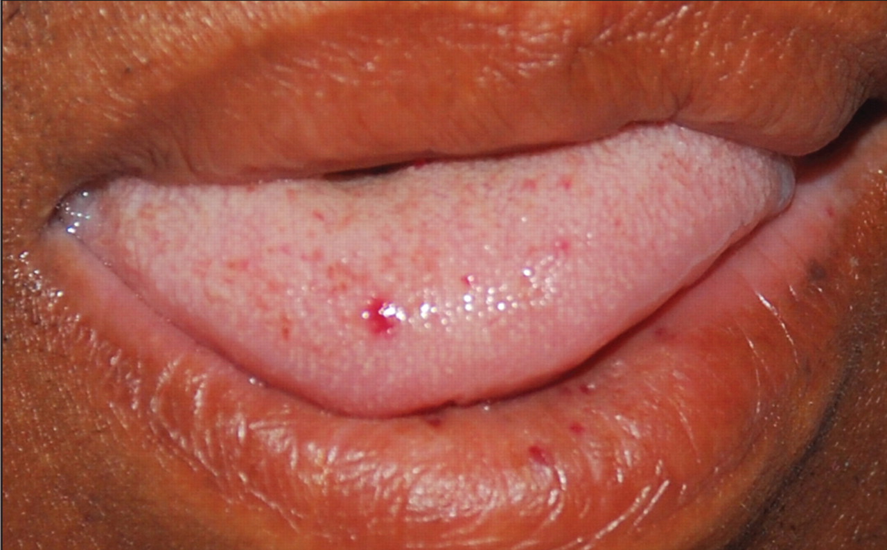
Figure 1: The patient had multiple telangiectasias on his tongue and lower lip.
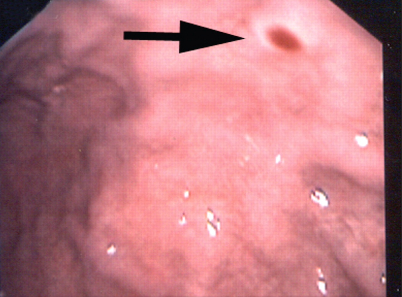
Figure 2: A gastric arteriovenous malformation surrounded by an anemic halo (arrow).
Figure 3:(A) A computed tomography scan of the patient’s abdomen (coronal view) showing a large hepatic arteriovenous malformation (arrow). (B) A 3-dimensional reconstructed image of the patient’s liver showing a large arteriovenous malformation (arrow).


In this issue
Article tools
Respond to this article


Related Articles
Cited By...
More in this TOC Section
Similar Articles

