
Main menu

  • Home
  • Content
    • Current issue
    • Past issues
    • Early releases
    • Collections
    • Sections
    • Blog
    • Infographics & illustrations
    • Podcasts
    • COVID-19 Articles
  • Authors & Reviewers
    • Overview for authors
    • Submission guidelines
    • Submit a manuscript
    • Forms
    • Editorial process
    • Editorial policies
    • Peer review process
    • Publication fees
    • Reprint requests
    • Open access
    • Patient engagement
  • Members & Subscribers
    • Benefits for CMA Members
    • CPD Credits for Members
    • Subscribe to CMAJ Print
    • Subscription Prices
  • Alerts
    • Email alerts
    • RSS
  • JAMC
    • À propos
    • Numéro en cours
    • Archives
    • Sections
    • Abonnement
    • Alertes
    • Trousse média 2023
  • CMAJ JOURNALS
    • CMAJ Open
    • CJS
    • JAMC
    • JPN

User menu

Search

  • Advanced search
CMAJ
  • CMAJ JOURNALS
    • CMAJ Open
    • CJS
    • JAMC
    • JPN
CMAJ

Advanced Search

  • Home
  • Content
    • Current issue
    • Past issues
    • Early releases
    • Collections
    • Sections
    • Blog
    • Infographics & illustrations
    • Podcasts
    • COVID-19 Articles
  • Authors & Reviewers
    • Overview for authors
    • Submission guidelines
    • Submit a manuscript
    • Forms
    • Editorial process
    • Editorial policies
    • Peer review process
    • Publication fees
    • Reprint requests
    • Open access
    • Patient engagement
  • Members & Subscribers
    • Benefits for CMA Members
    • CPD Credits for Members
    • Subscribe to CMAJ Print
    • Subscription Prices
  • Alerts
    • Email alerts
    • RSS
  • JAMC
    • À propos
    • Numéro en cours
    • Archives
    • Sections
    • Abonnement
    • Alertes
    • Trousse média 2023
  • Visit CMAJ on Facebook
  • Follow CMAJ on Twitter
  • Follow CMAJ on Pinterest
  • Follow CMAJ on Youtube
  • Follow CMAJ on Instagram
Practice

A spontaneous skin lesion

Josie McCabe, Zoe Blades and Emmet E. McGrath
CMAJ December 02, 2008 179 (12) 1297-1299; DOI: https://doi.org/10.1503/cmaj.080635
Josie McCabe MB
  • Find this author on Google Scholar
  • Find this author on PubMed
  • Search for this author on this site
Zoe Blades MB
  • Find this author on Google Scholar
  • Find this author on PubMed
  • Search for this author on this site
Emmet E. McGrath MB
  • Find this author on Google Scholar
  • Find this author on PubMed
  • Search for this author on this site
  • Article
  • Figures & Tables
  • Responses
  • Metrics
  • PDF
Loading
  • © 2008 Canadian Medical Association

A 34-year-old woman presented with long-standing severe asthma and chronic idiopathic angioedema. We found 2 lesions on her back (Figure 1, Figure 2). The lesions had occurred spontaneously 10 years ago and had grown slightly bigger over that time. They were quite painful, tender and itchy at times. The patient had no history of trauma, surgery or acne. She was seen by a dermatologist for further assessment and continues to attend the outpatient department.

Figure
  • Download figure
  • Open in new tab
  • Download powerpoint

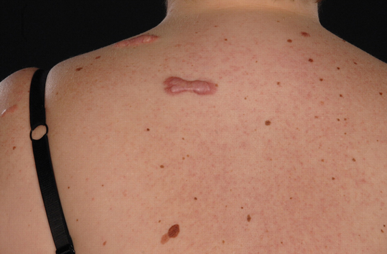
Figure 1: Irregularly shaped, raised pink lesions on the upper back of a 34-year-old woman.

Figure
  • Download figure
  • Open in new tab
  • Download powerpoint

Figure 2: A close-up image shows the irregular margins and smooth, shiny surface of the lesions.

    What is your diagnosis?

  1. Cutaneous sarcoidosis

  2. Keloid

  3. Dermatofibroma

  4. Dermatofibrosarcoma protuberans

  5. Hypertrophic scar

    What might be a reasonable next step in treating this patient?

  1. Biopsy to confirm your diagnosis

  2. Prescription of topical high-potency steroid cream

  3. Intralesional steroid injection

  4. Surgical excision of the lesion

  5. HLA genotyping of the patient

Discussion

The diagnosis is (b) keloid, and the next step in treatment is (c) intralesional steroid injection. The patient was given the intralesional steroid injections, with occlusive dressings and silicone gel. No change was seen in the keloid.

Keloid is a condition characterized by excessive deposition of fibrous tissue. It usually develops after trauma or injury (e.g., earlobe piercing, surgery, acne, chickenpox, surgical incision, burns or vaccination).1 The diagnosis of our patient's condition was straightforward because of the classic nature of the findings: a firm nodule on the upper back that was present for a long time, was relatively unchanged, had a smooth surface and a claw-like, skin-coloured appearance, and was prone to itchiness and tenderness.

The texture of keloids ranges from soft and doughy to rubbery and hard. Keloids vary in colour with age, from erythematous through brownish red to pale pink. Keloidal collagen is arranged haphazardly instead of parallel to the skin surface, as in normal scar formation.2

Keloids grow in a claw-like fashion outward in all directions from the original wound. The extent of growth is grossly out of proportion to the degree of tissue damage. The condition is more prevalent in women than in men. It tends to occur in people aged 10–30 years. Rarely, it can occur in older people (e.g., patients undergoing coronary artery bypass surgery).

People of Polynesian and Chinese backgrounds are most susceptible to keloids, whereas white people are least commonly affected.1 Genetic associations have been identified with several HLA genotypes, including HLA-B14, HLA-B21, HLA-Bw16, HLA-DR5, HLA-DQw3 and blood group A.1

Spontaneous keloid formation is rare. Whether it is truly spontaneous is controversial. The scar tissue may form after an insignificant inflammatory reaction or injury — one that was too trivial for the person to remember. Confirmed instances of spontaneous keloid have been reported in Dubowitz syndrome, Rubenstein–Taybi syndrome and Noonan syndrome.3 Spontaneous keloid has also been reported in siblings3 and in people with allergic disease.4

The differential diagnosis of keloid includes hypertrophic scar, dermatofibroma, dermatofibrosarcoma protuberans and cutaneous sarcoidosis (Table 1). Unlike keloid, hypertrophic scar never occurs spontaneously or extends beyond the margins of the scar. We excluded dermatofibroma because our patient's lesions were neither coloured nor very small or located on the extremities. We also excluded dermatofibrosarcoma protuberans because the lesions did not invade the deeper structures, had not increased considerably in size over time and had not ulcerated. Cutaneous sarcoidosis, known as the great imitator, presents with a variety of appearances, including that of a raised nodular lesion. Its diagnosis requires a biopsy demonstrating non-caseating granuloma. Since the classic characteristics of keloid were present, we did not perform a biopsy, which could have worsened the scar. Biopsy would be a reasonable next step if clinical suspicion or other investigations pointed to another diagnosis (e.g., sarcoidosis).

View this table:
  • View inline
  • View popup
  • Download powerpoint

Table 1.

The optimal treatment for keloid depends on several factors, including previous response to treatment, the patient's age, and the location and size of the affected area. Prevention is better than cure. The “gold standard” treatment is excision followed by careful postoperative care (using occlusive dressings, compression therapy, intralesional corticosteroid injections or radiotherapy). Excision alone should be avoided, because it is associated with high recurrence rates.2 Other treatment options include laser ablation, cryotherapy and drug therapy such as interferons, tamoxifen, verapamil and retinoic acid.1,2

Although many treatment options exist, no treatment alone has proven effective. Most patients experience recurrence following treatment.

Footnotes

  • CMAJ invites submissions to “What is your call?” Clinical details (including images) are presented on the first page along with a multiple-choice question about the diagnosis. The answer and a brief discussion of the condition follow on the second page. We specifically invite submissions illustrating common or important radiographic and electrocardiographic diagnoses of appeal to a general audience. We allow up to 5 references and require authors to obtain consent from the patient for publication of his or her story (form available at www.cmaj.ca/authors/checklist.shtml). Submit manuscripts online at http://mc.manuscriptcentral.com/cmaj.

    This article has been peer reviewed.

    Competing interests: None declared.

REFERENCES

  1. 1.↵
    Berman B, Zell D, Perez OA. Keloid and hypertrophic scar. WebMD emedicine 2007 Feb 1. Available: www.emedicine.com/DERM/topic205.htm (accessed 2008 Oct 2).
  2. 2.↵
    Berman B, Flores F. The treatment of hypertrophic scars and keloids. Eur J Dermatol 1998;8:591-5.
    OpenUrlPubMed
  3. 3.↵
    Mandal A, Imran D, Rao GS. Spontaneous keloids in siblings. Ir Med J 2004;97:250-1.
    OpenUrlPubMed
  4. 4.↵
    Oittinen HA, O'Shaughnessy M. Multiple nonsyndromic spontaneous keloids in allergic disease. Plast Reconstr Surg 2007;119:762-3.
    OpenUrlCrossRefPubMed
  5. 5.
    Naversen DN, Trask DM, Watson FH, et al. Painful tumors of the skin: “LEND AN EGG”. J Am Acad Dermatol 1993;28(2 pt 2):298-300.
    OpenUrlPubMed
PreviousNext
Back to top

In this issue

Canadian Medical Association Journal: 179 (12)
CMAJ
Vol. 179, Issue 12
2 Dec 2008
  • Table of Contents
  • Index by author

Article tools

Respond to this article
Print
Download PDF
Article Alerts
To sign up for email alerts or to access your current email alerts, enter your email address below:
Email Article

Thank you for your interest in spreading the word on CMAJ.

NOTE: We only request your email address so that the person you are recommending the page to knows that you wanted them to see it, and that it is not junk mail. We do not capture any email address.

Enter multiple addresses on separate lines or separate them with commas.
A spontaneous skin lesion
(Your Name) has sent you a message from CMAJ
(Your Name) thought you would like to see the CMAJ web site.
CAPTCHA
This question is for testing whether or not you are a human visitor and to prevent automated spam submissions.
Citation Tools
A spontaneous skin lesion
Josie McCabe, Zoe Blades, Emmet E. McGrath
CMAJ Dec 2008, 179 (12) 1297-1299; DOI: 10.1503/cmaj.080635

Citation Manager Formats

  • BibTeX
  • Bookends
  • EasyBib
  • EndNote (tagged)
  • EndNote 8 (xml)
  • Medlars
  • Mendeley
  • Papers
  • RefWorks Tagged
  • Ref Manager
  • RIS
  • Zotero
‍ Request Permissions
Share
A spontaneous skin lesion
Josie McCabe, Zoe Blades, Emmet E. McGrath
CMAJ Dec 2008, 179 (12) 1297-1299; DOI: 10.1503/cmaj.080635
Digg logo Reddit logo Twitter logo Facebook logo Google logo Mendeley logo
  • Tweet Widget
  • Facebook Like

Jump to section

  • Article
    • Discussion
    • Footnotes
    • REFERENCES
  • Figures & Tables
  • Responses
  • Metrics
  • PDF

Related Articles

  • Highlights
  • Dans ce numéro
  • PubMed
  • Google Scholar

Cited By...

  • A Rare Case of Postauricular Spontaneous Keloid in an Elderly Patient
  • Google Scholar

More in this TOC Section

  • A blistering variant of phlegmasia cerulea dolens from underlying squamous cell lung cancer
  • Parechovirus infections in infants
  • Radiation dermatitis in a patient treated for hepatocarcinoma
Show more Practice

Similar Articles

Collections

  • Topics
    • Dermatology


View Latest Classified Ads

Content

  • Current issue
  • Past issues
  • Collections
  • Sections
  • Blog
  • Podcasts
  • Alerts
  • RSS
  • Early releases

Information for

  • Advertisers
  • Authors
  • Reviewers
  • CMA Members
  • CPD credits
  • Media
  • Reprint requests
  • Subscribers

About

  • General Information
  • Journal staff
  • Editorial Board
  • Advisory Panels
  • Governance Council
  • Journal Oversight
  • Careers
  • Contact
  • Copyright and Permissions
  • Accessibiity
  • CMA Civility Standards
CMAJ Group

Copyright 2023, CMA Impact Inc. or its licensors. All rights reserved. ISSN 1488-2329 (e) 0820-3946 (p)

All editorial matter in CMAJ represents the opinions of the authors and not necessarily those of the Canadian Medical Association or its subsidiaries.

To receive any of these resources in an accessible format, please contact us at CMAJ Group, 500-1410 Blair Towers Place, Ottawa ON, K1J 9B9; p: 1-888-855-2555; e: cmajgroup@cmaj.ca

Powered by HighWire