
Main menu

  • Home
  • COVID-19
    • Articles & podcasts
    • Blog posts
    • Collection
    • News
  • Content
    • Current issue
    • Past issues
    • Early releases
    • Collections
    • Sections
    • Blog
    • Infographics & illustrations
    • Podcasts
    • Classified ads
  • Authors
    • Overview for authors
    • Submission guidelines
    • Submit a manuscript
    • Forms
    • Editorial process
    • Editorial policies
    • Peer review process
    • Publication fees
    • Reprint requests
  • CMA Members
    • Overview for members
    • Earn CPD Credits
    • Print copies of CMAJ
    • Career Ad Discount
  • Subscribers
    • General information
    • View prices
  • Alerts
    • Email alerts
    • RSS
  • JAMC
    • À propos
    • Numéro en cours
    • Archives
    • Sections
    • Abonnement
    • Alertes
  • CMAJ JOURNALS
    • CMAJ Open
    • CJS
    • JAMC
    • JPN

User menu

Search

  • Advanced search
CMAJ
  • CMAJ JOURNALS
    • CMAJ Open
    • CJS
    • JAMC
    • JPN
CMAJ

Advanced Search

  • Home
  • COVID-19
    • Articles & podcasts
    • Blog posts
    • Collection
    • News
  • Content
    • Current issue
    • Past issues
    • Early releases
    • Collections
    • Sections
    • Blog
    • Infographics & illustrations
    • Podcasts
    • Classified ads
  • Authors
    • Overview for authors
    • Submission guidelines
    • Submit a manuscript
    • Forms
    • Editorial process
    • Editorial policies
    • Peer review process
    • Publication fees
    • Reprint requests
  • CMA Members
    • Overview for members
    • Earn CPD Credits
    • Print copies of CMAJ
    • Career Ad Discount
  • Subscribers
    • General information
    • View prices
  • Alerts
    • Email alerts
    • RSS
  • JAMC
    • À propos
    • Numéro en cours
    • Archives
    • Sections
    • Abonnement
    • Alertes
  • Visit CMAJ on Facebook
  • Follow CMAJ on Twitter
  • Follow CMAJ on Pinterest
  • Follow CMAJ on Youtube
  • Follow CMAJ on Instagram
Practice

Widened mediastinum in a child with severe trauma

Ilan Buffo-Sequeira and Douglas D. Fraser
CMAJ November 06, 2007 177 (10) 1181-1182; DOI: https://doi.org/10.1503/cmaj.070936
Ilan Buffo-Sequeira MD
  • Find this author on Google Scholar
  • Find this author on PubMed
  • Search for this author on this site
Douglas D. Fraser MD PhD
  • Find this author on Google Scholar
  • Find this author on PubMed
  • Search for this author on this site
  • Article
  • Figures & Tables
  • Responses
  • Metrics
  • PDF
Loading
  • © 2007 Canadian Medical Association

A 3-year-old girl was struck by a vehicle travelling at a high speed and was thrown approximately 6 m. Emergency medical services staff noted that the patient was seizing with left-deviated eyes. She was rushed to the closest community hospital for resuscitation and was subsequently transferred to our regional trauma centre.

Despite administration of about 55 mL of crystalloid per kilogram body weight, the patient was tachycardic and hypotensive for her age. A trauma blood panel was drawn, and noncrossmatched blood was transfused. Laboratory test results suggested occult bleeding and myocardial contusion (hemoglobin concentration 81 [normal 110–160] g/L, hematocrit 0.24 [normal 0.33–0.48] and troponin I 0.39 [normal < 0.08] μg/L).

Figure1
  • Download figure
  • Open in new tab
  • Download powerpoint

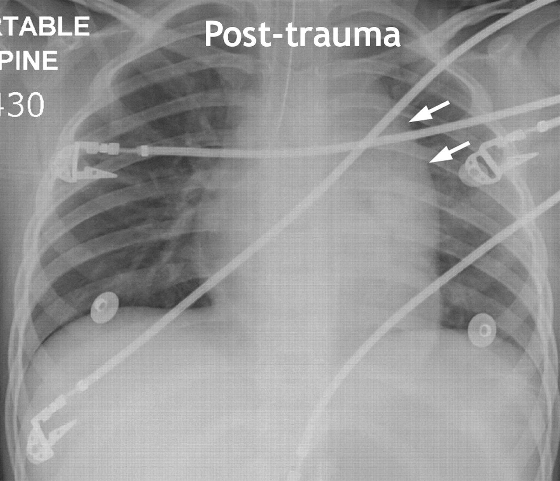
Figure 1: Chest radiograph (anteroposterior) taken several hours following the trauma, showing an apparent widened mediastinum with loss of the aortic knob contour (arrows). Deviation of the esophagus could not be excluded because a nonopaque orogastric tube had been inserted before transfer to the trauma centre.

A chest radiograph taken with the patient in a supine position showed a widened mediastinum with loss of the aortic knob contour (Figure 1). Although a frank apical cap or hemothorax was absent, a small amount of fluid was present in the left pleural apex. The left main bronchus was in normal anatomic position. A normal chest radiograph with the patient in an upright position had been taken several months before the trauma and was available for comparison (Figure 2).

Figure2
  • Download figure
  • Open in new tab
  • Download powerpoint

Figure 2: Normal chest radiograph (posteroanterior) with patient in upright position, taken several months before the trauma.

Once the patient's condition was stabilized, contrast-enhanced computed tomography was performed and showed a normal aorta with no evidence of hematoma (Figure 3). The thymus accounted for the apparent widened mediastinum seen on the chest radiograph. Intracranial bleeding, identified on a computed tomography scan of her head, accounted for the blood loss. The patient survived and is currently managed at a neurorehabilitation centre.

Figure3
  • Download figure
  • Open in new tab
  • Download powerpoint

Figure 3: Computed tomography scan, showing normal mediastinum with no evidence of aortic filling defects or pseudoaneurysm. A large thymus (asterisks) consistent with age is evident. A mediastinal fat density (arrow) and pleural effusion (double arrow) are present.

A widened mediastinum in trauma patients should be considered an aortic injury until proven otherwise. In children, traumatic aortic injuries are uncommon (in about 0.1% of cases) because of the relative elasticity of their great vessels compared with those in adults. When aortic injury occurs in a child it is most commonly seen at the level of the ligamentum arteriosus and is usually associated with severe deceleration forces.1 The signs of aortic injury on a chest radiograph include mediastinal widening, loss of the aortic knob contour, depression of the left main bronchus, deviation of the trachea or esophagus, left pleural cap, hemothorax and fracture of the first or second rib.1,2

As illustrated in this case, interpretation of the medistinal outline on a chest radiograph in younger children in the supine position is complicated by the presence of the thymus.3 A chest radiograph taken with the child in an upright position can be obtained to clarify the medistinal borders; however, placing a trauma patient with unknown injuries in such a position poses an unacceptable risk. Although arch aortography remains the “gold standard,” contrast-enhanced computed tomography and transesophageal echocardiography are acceptable methods to exclude aortic injuries.1–3 Computed tomography scans of the chest have the added advantage of delineating the thymic outline.1

Footnotes

  • This article has been peer reviewed.

    Competing interests: None declared.

REFERENCES

  1. 1.↵
    Bliss D, Silen M. Pediatric thoracic trauma. Crit Care Med 2002;30:S409-15.
  2. 2.↵
    Stafford PW, Blinman TA, Nance ML. Practical points in evaluation and resuscitation of the injured child. Surg Clin North Am 2002;82:273-301.
    OpenUrlCrossRefPubMed
  3. 3.↵
    Mirvis SE, Shanmuganathan K, editors. Imaging in trauma and critical care. 2nd ed. Philadelphia: Saunders/Elsevier Science; 2003. p. 711–24.
PreviousNext
Back to top

In this issue

Canadian Medical Association Journal: 177 (10)
CMAJ
Vol. 177, Issue 10
6 Nov 2007
  • Table of Contents
  • Index by author

Article tools

Respond to this article
Print
Download PDF
Article Alerts
To sign up for email alerts or to access your current email alerts, enter your email address below:
Email Article

Thank you for your interest in spreading the word on CMAJ.

NOTE: We only request your email address so that the person you are recommending the page to knows that you wanted them to see it, and that it is not junk mail. We do not capture any email address.

Enter multiple addresses on separate lines or separate them with commas.
Widened mediastinum in a child with severe trauma
(Your Name) has sent you a message from CMAJ
(Your Name) thought you would like to see the CMAJ web site.
CAPTCHA
This question is for testing whether or not you are a human visitor and to prevent automated spam submissions.
Citation Tools
Widened mediastinum in a child with severe trauma
Ilan Buffo-Sequeira, Douglas D. Fraser
CMAJ Nov 2007, 177 (10) 1181-1182; DOI: 10.1503/cmaj.070936

Citation Manager Formats

  • BibTeX
  • Bookends
  • EasyBib
  • EndNote (tagged)
  • EndNote 8 (xml)
  • Medlars
  • Mendeley
  • Papers
  • RefWorks Tagged
  • Ref Manager
  • RIS
  • Zotero
‍ Request Permissions
Share
Widened mediastinum in a child with severe trauma
Ilan Buffo-Sequeira, Douglas D. Fraser
CMAJ Nov 2007, 177 (10) 1181-1182; DOI: 10.1503/cmaj.070936
Digg logo Reddit logo Twitter logo Facebook logo Google logo Mendeley logo
  • Tweet Widget
  • Facebook Like
  • Google Plus One

Jump to section

  • Article
    • Footnotes
    • REFERENCES
  • Figures & Tables
  • Responses
  • Metrics
  • PDF

Related Articles

  • Highlights of this issue
  • Dans ce numéro
  • PubMed
  • Google Scholar

Cited By...

  • No citing articles found.
  • Google Scholar

More in this TOC Section

  • “Superscan” in diffusion-weighted imaging with background body suppression magnetic resonance imaging
  • Using intranasal corticosteroids
  • Langerhans cell histiocytosis in a 5-month-old baby
Show more Practice

Similar Articles

Collections

  • Topics
    • Trauma, injury & trauma surgery
    • Pediatrics

Content

  • Current issue
  • Past issues
  • Collections
  • Sections
  • Blog
  • Podcasts
  • Alerts
  • RSS
  • Early releases

Information for

  • Advertisers
  • Authors
  • Reviewers
  • CMA Members
  • Media
  • Reprint requests
  • Subscribers

About

  • General Information
  • Journal staff
  • Editorial Board
  • Governance Council
  • Journal Oversight
  • Careers
  • Contact
  • Copyright and Permissions

Copyright 2021, Joule Inc. or its licensors. All rights reserved. ISSN 1488-2329 (e) 0820-3946 (p)

All editorial matter in CMAJ represents the opinions of the authors and not necessarily those of the Canadian Medical Association or its subsidiaries.

Powered by HighWire