


- © 2004 Canadian Medical Association or its licensors
A previously well 60-year-old woman presented to hospital with a 3-week history of fatigue, anorexia, abdominal pain, nausea and vomiting, and a 4-kg weight loss. She had previously consulted her general practitioner about an episode of diarrhea and dark stools and was found at that time to have anemia (hemoglobin level 67 g/L, mean corpuscular volume 65.1 fL) with mild leukocytosis (leukocyte count 16.6 х109/L, neutrophil count 13.2 х109/L) and thrombocytosis (platelet count 975 х109/L). Her history included postpolio syndrome, retinitis pigmentosa, benign nontoxic goiter (for which she was taking thyroxine as suppressive therapy) and, 11 years before admission, excision of a nodular melanoma of the skin (Clark level V: penetration through the dermis to the subcutaneous fat).
On examination the woman was found to have a firm, irregular, mobile mass about the size of a tennis ball in her left lower quadrant and tenderness, but no rebound or guarding. Her bowel sounds were hyperactive. The woman was admitted to hospital for bowel rest, intravenous fluids and investigation of her anemia.
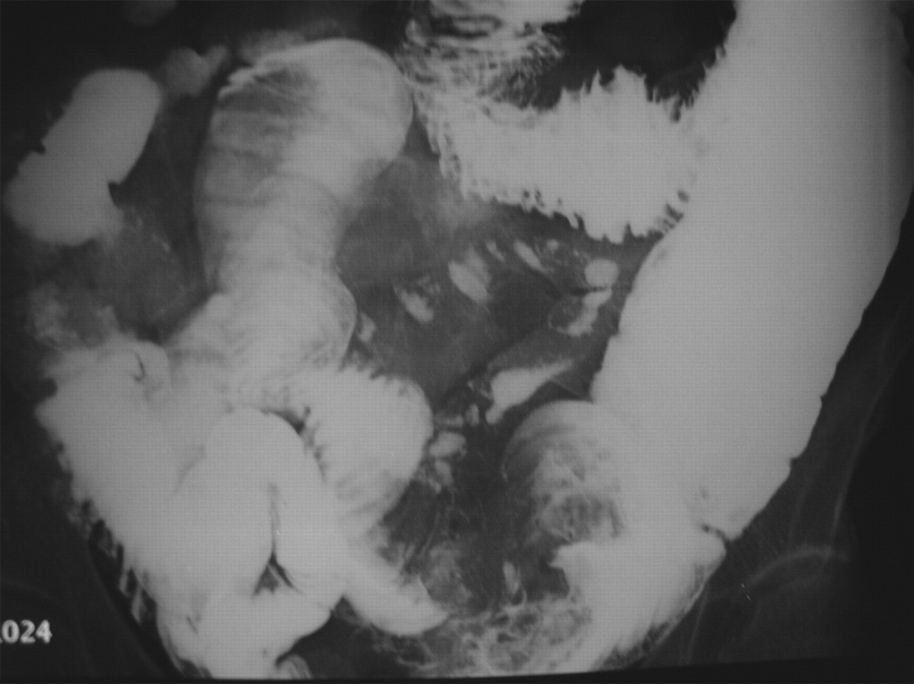
Three separate stool specimens were positive for occult blood. Colonoscopy revealed uncomplicated diverticulosis, and results of upper endoscopy and mucosal biopsies were unremarkable. A small-bowel follow-through series showed a mass lesion in the lower left quadrant, with a dilated proximal jejunal loop (Fig. 1). At open laparotomy, a single, large, 9-cm intramural nodule with an ulcerated mucosa was found in the patient's jejunum. The mass was resected and identified histologically and immunochemically to be a melanoma metastatic to the small intestine. Postoperatively, the woman's gastrointestinal symptoms resolved, and she was discharged home. Despite 2 small lung nodules identified on a subsequent high-resolution CT scan of the chest, the patient remained well 8 months after being admitted. She declined interferon-α treatment.
Melanoma is the most common tumour to metastasize to the gastrointestinal tract, in particular the small intestine, where it can simulate acute appendicitis and cause gastrointestinal bleeding or obstruction.1 Although 4% of patients with melanoma are found to have clinically significant metastases to the small intestine, many more such metastases are identified at autopsy.2 A recent case series included 32 melanoma patients with confirmed metastasis to the small intestine.3 The most common presenting symptom was abdominal pain (60% of patients); other symptoms included bowel obstruction (47%), nausea and vomiting (41%) and gastrointestinal bleeding (30%). An abdominal mass was identified at presentation in only 10% of patients.3 Given the relatively low sensitivity of imaging studies (from 58% for small-bowel follow-through exams to 66% for contrast-enhanced CT scans),3 patients with a history of melanoma who have unexplained abdominal pain or anemia, or both, often undergo exploratory surgery to rule out gastrointestinal metastases. About 50% with gastrointestinal metastases have 3 or fewer lesions.3 Although the textbook description of the gross appearance of these lesions is one of a discrete polypoid mass with a central ulceration (a “bull's eye” sign),4 such an appearance is uncommon. In the absence of other obvious distant metastases, tumours are often resected surgically. Resection allows a definitive diagnosis and, although a cure is rare, can prolong overall and symptom-free survival.5
Mario Pirisi Monica Leutner Sandra Grazioli Ettore G. Bartoli Department of Medical Sciences University of East Piedmont Novara, Italy
In this issue

Article tools


Jump to section
Related Articles
Cited By...
- No citing articles found.
More in this TOC Section
Similar Articles
Collections

