


- © 2004 Canadian Medical Association or its licensors
Fig. 1: Life cycle of the guinea worm. Photo: Chesley Sheppard
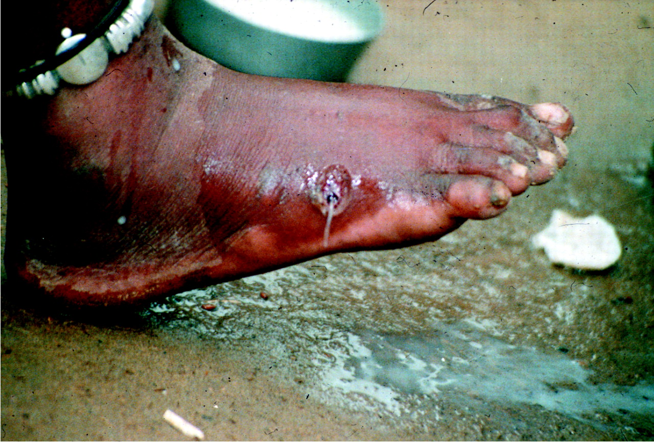
Fig. 3: Guinea worm emerging from foot ulcer. Photo: The Carter Center / S. Fitzgerald
Fig. 2: Foot blister induced by the female guinea worm in a person with dracunculiasis (guinea worm disease). Photo: The Carter Center / S. Fitzgerald
Fig. 4: Map of current and former dracunculiasis-endemic countries, 2002. Photo: WHO Collaborating Center for Research, Training and Eradication of Dracunculiasis
Fig. 6: A young man with guinea worm disease receives education on the life cycle of the guinea worm and how to prevent contamination of drinking-water sources. Photo: The Carter Center / Emily Howard
Fig. 5: A Togolese woman strains her family's drinking water through a cloth filter to prevent them from contracting guinea worm disease. Photo: The Carter Center / Emily Howard


- © 2004 Canadian Medical Association or its licensors
In this issue
Article tools


Jump to section
Related Articles
Cited By...
- No citing articles found.
More in this TOC Section
Similar Articles
Collections

