


Abstract
CONGENITAL VARICELLA SYNDROME refers to the spectrum of fetal anomalies associated with maternal varicella zoster virus (VZV) infection during the first trimester of pregnancy. The syndrome is rare and the risk to the fetus uncertain. We describe an unusual case of congenital varicella syndrome in which hydrocephalus was the main consequence and likely represented VZV reactivation in utero.
Case report
A male infant was delivered by means of cesarean section after 37 weeks' gestation. The mother, a 29-year-old gravida 2, para 1 woman, was exposed to varicella zoster virus (VZV) during week 15. Varicella infection was diagnosed on clinical grounds following a classic presentation of chickenpox. No antiviral treatment was given. Ultrasound examination 12 weeks later revealed symmetrically impaired fetal growth and bilateral ventriculomegaly.
At birth the infant weighed 2190 g (below the 10th percentile) and was 42 cm long (also below the 10th percentile). The Apgar score was 5 and 6 at 1 minute and 5 minutes respectively. Endotracheal intubation was performed at 7 minutes because of the infant's erratic respiratory drive. Physical examination revealed severe hydrocephalus (head circumference 36.5 cm [above the 90th percentile]), large anterior fontanelle (4 х 4 cm), diastasis of the sagittal suture, asymmetry of the rima oris, severe microphthalmos of the left eye, and cutaneous scars following a dermatomal distribution on the left side of the head and neck, the left ear and the right lower limb (Fig. 1). There was also decreased physical activity and diminished muscle tone. Tonic-clonic seizures began 4 hours after birth and were treated with phenobarbital. Ultrasound examination confirmed bilateral ventriculomegaly.
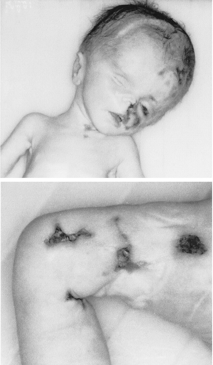
Fig. 1: Gross appearance of patient showing hydrocephalus, left-sided microphthalmia and cutaneous ulceration involving the head, neck and right lower limb.
Treatment with acyclovir (30 mg/kg daily in 3 divided doses) was begun on the first day of life. Despite intensive care and respiratory support, the infant's clinical course rapidly deteriorated, and he died on the third day of life.
Antemortem and postmortem cultures of both the cerebrospinal fluid (CSF) and blood were negative for bacteria. Analysis of the CSF revealed pleocytosis, elevated protein levels and a low glucose level. VZV IgG antibodies were present at a titre of 12 IU in the mother and 11 IU in the infant; IgM antibodies were not present in either. VZV DNA was detected by means of polymerase chain reaction testing in the CSF specimen. Antibodies to herpes simplex, cytomegalovirus, rubella and Toxoplasma sp. were absent. Chromosome analysis showed a 46 XY karyotype.
At autopsy the most severe lesions were found in the central nervous system. Gross examination revealed marked triventricular noncommunicating hydrocephalus with little remaining cortex. The aqueduct of Sylvius was occluded by an intense gliosis. The only structures that appeared normal were the basal ganglia and thalamus. Microscopically the grey matter showed neuronal loss and massive mononuclear infiltration, with a prominent reaction by microglial clusters, microcalcification and a perivascular accumulation of mononuclear cells (mainly lymphocytes and monocytes) (Fig. 2, panel A). Most of the lesions in the white matter appeared as necrotic areas with macrophages and focal hemorrhage surrounded by a paucicellular and edematous periphery of myelin loss rimmed by many reactive astrocytes. Periventricular congestion and hemorrhage indicated ependymitis, with focal discontinuities of the cellular lining (Fig. 2, panel B). There was perivascular cuffing of the subependymal small vessels by lymphocytes (Fig. 2, panel C). The internal carotid arteries and the major cerebral arteries were normal. There was evidence of neuronal loss and mononuclear infiltration in the left trigeminal ganglia.

Fig. 2: Panel A: Necrotizing encephalitis with varying degrees of gliosis and mononuclear infiltration by lymphocytes and plasma cells (hematoxylin–eosin, original magnification х10). Panel B: Focal discontinuities of the ependymal lining with subependymal infiltration by lymphocytes and gliosis (hematoxylin–eosin, original magnification х10). Panel C: Perivascular cuffing of the subependyma vessels by lymphocytes (hematoxylin–eosin, original magnification х25).
Evaluation of the left orbital contents revealed an extremely small and malformed globe, with only rudimentary ocular contents. The optic nerve appeared severely hypoplastic. The right eye appeared normal on gross and microscopic examination.
On gross examination the lungs were found to be diffusely congested and covered by hemorrhagic pleura. Histologic examination showed interstitial mononuclear infiltration surrounding the small and large airways, with intra-alveolar proteinaceous exudate.
Full-thickness sections of epidermal ulcers showed large areas of spongiosis, with minimal inflammation.
Comments
Congenital varicella syndrome (CVS) is a rare disorder resulting from maternal–fetal VZV transmission, usually between 8 and 20 weeks' gestation.1,2 The incidence of maternal VZV infection has been estimated to be between 5 and 7 cases per 10 000 pregnancies.2 Fetus-specific factors play a crucial role, and fetal consequences of maternal exposure may be highly variable.3 Given that up to 2% of fetuses infected between 8 and 20 weeks' gestation may exhibit pathogenetic effects of VZV,4 we estimate the CVS incidence rate to be about 2.8–4 cases per 100 000 pregnancies.
Since the description of CVS by Laforet and Lynch in 1947,5 embryopathy following fetal infection with VZV has been well described in case reports.1,2,6 More recently, a number of reports have focused on the complications of VZV reactivation.7,8 Although zoster infection may develop in the neonatal period, descriptions of intrauterine reactivation are very rarely reported.
CVS manifests as cutaneous scars, limb hypoplasia, muscle atrophy, malformation of the digits, psychomotor retardation, microcephaly, cortical atrophy, Horner's syndrome and various eye abnormalities, including cataracts, chorioretinitis and microphthalmos. Intrauterine growth retardation as well as motor and sensory deficits have also been described.1,2
The case we have presented exhibited most of the clinical patterns of CVS, namely smallness for date, dermatomal distribution of skin lesions and ocular abnormalities, but our patient also had hydrocephalus, a very unusual stigma. We were able to find only 1 article about CVS in which hydrocephalus was reported (in 1 of 9 cases of congenital varicella).9 A recent review of the literature failed to show hydrocephalus among clinical findings in 77 infants whose mothers had had VZV infection during pregnancy.1
Undoubtedly, VZV has a well-known teratogenic role in embryopathy. The abnormalities of CVS appear to be secondary to the denervation of fetal structures as a result of the neurotropic nature of the virus and of exposure during embryogenesis.10 However, in our case the dermatomal distribution of the skin lesions, the histopathological finding of neuronal loss and mononuclear infiltration in the sensory ganglia, together with the presence of VZV DNA in the CSF and evidence of encephalitis, strongly support the probability of an intrauterine reactivation of the virus following the primary infection.
Although the biological mechanisms underlying the transition from latency to active viral replication are still unknown, the traditional concept of VZV reactivation requires the replication and spread of latent virus in the ganglion cells and the central nervous system.1,7
Recently, renewed interest has focused on neurological complications of VZV reactivation.7,8 Among them, encephalitis caused by small-vessel vasculitis with predominant ventriculitis, periventricular dilatation and consequent hydrocephalus is a well-known but unusual presentation in immunocompromised adults.7,11 The pathological changes we found, namely the perivascular cuffing of the subependymal small vessels by lymphocytes and ependymitis, are consistent with this picture.
On the basis of these observations, we hypothize that, in our patient, VZV reactivation occurred after the primary infection, and the zoster infection spread to the spinal cord and brain. Hydrocephalus may have developed as a consequence of ependymitis owing to blockage of CSF outflow at the level of the aqueduct of Sylvius. Cortical destruction resulted from both involvement of the penetrating vessels and pressure atrophy. The brevity of the latent period between primary infection and reactivation was most likely due to the fetus' immature immune system,12,13 which would also explain the extensive damage seen in the developing fetus as compared with damage caused by zoster infection in adults.
Footnotes
-
This article has been peer reviewed.
Contributors: All authors contributed substantially to the conception and design of the article. In addition, each author revised the paper critically and approved the final version.
Competing interests: None declared.
In this issue
Article tools


Jump to section
Related Articles
Cited By...
More in this TOC Section
Research
Similar Articles
Collections

