
Main menu

  • Home
  • COVID-19
    • Articles & podcasts
    • Blog posts
    • Collection
    • News
  • Content
    • Current issue
    • Past issues
    • Early releases
    • Collections
    • Sections
    • Blog
    • Infographics & illustrations
    • Podcasts
  • Authors
    • Overview for authors
    • Submission guidelines
    • Submit a manuscript
    • Forms
    • Editorial process
    • Editorial policies
    • Peer review process
    • Publication fees
    • Reprint requests
  • CMA Members
    • Overview for members
    • Earn CPD Credits
    • Print copies of CMAJ
    • Career Ad Discount
  • Subscribers
    • General information
    • View prices
  • Alerts
    • Email alerts
    • RSS
  • JAMC
    • À propos
    • Numéro en cours
    • Archives
    • Sections
    • Abonnement
    • Alertes
    • Trousse média 2021
  • CMAJ JOURNALS
    • CMAJ Open
    • CJS
    • JAMC
    • JPN

User menu

Search

  • Advanced search
CMAJ
  • CMAJ JOURNALS
    • CMAJ Open
    • CJS
    • JAMC
    • JPN
CMAJ

Advanced Search

  • Home
  • COVID-19
    • Articles & podcasts
    • Blog posts
    • Collection
    • News
  • Content
    • Current issue
    • Past issues
    • Early releases
    • Collections
    • Sections
    • Blog
    • Infographics & illustrations
    • Podcasts
  • Authors
    • Overview for authors
    • Submission guidelines
    • Submit a manuscript
    • Forms
    • Editorial process
    • Editorial policies
    • Peer review process
    • Publication fees
    • Reprint requests
  • CMA Members
    • Overview for members
    • Earn CPD Credits
    • Print copies of CMAJ
    • Career Ad Discount
  • Subscribers
    • General information
    • View prices
  • Alerts
    • Email alerts
    • RSS
  • JAMC
    • À propos
    • Numéro en cours
    • Archives
    • Sections
    • Abonnement
    • Alertes
    • Trousse média 2021
  • Visit CMAJ on Facebook
  • Follow CMAJ on Twitter
  • Follow CMAJ on Pinterest
  • Follow CMAJ on Youtube
  • Follow CMAJ on Instagram
Clinical Vistas

A rare site for tuberculosis

Amitabh Monga, Anju Arora, Ravinder Pal Singh Makkar and Ajay K. Gupta
CMAJ November 12, 2002 167 (10) 1149-1150;
Amitabh Monga
Department of Internal Medicine Sitaram Bhartia Institute of Science and ResearchNew Delhi, India
  • Find this author on Google Scholar
  • Find this author on PubMed
  • Search for this author on this site
Anju Arora
Department of Internal Medicine Sitaram Bhartia Institute of Science and ResearchNew Delhi, India
  • Find this author on Google Scholar
  • Find this author on PubMed
  • Search for this author on this site
Ravinder Pal Singh Makkar
Department of Internal Medicine Sitaram Bhartia Institute of Science and ResearchNew Delhi, India
  • Find this author on Google Scholar
  • Find this author on PubMed
  • Search for this author on this site
Ajay K. Gupta
Department of Internal Medicine Sitaram Bhartia Institute of Science and ResearchNew Delhi, India
  • Find this author on Google Scholar
  • Find this author on PubMed
  • Search for this author on this site
  • Article
  • Figures & Tables
  • Metrics
  • Responses
  • PDF
Loading

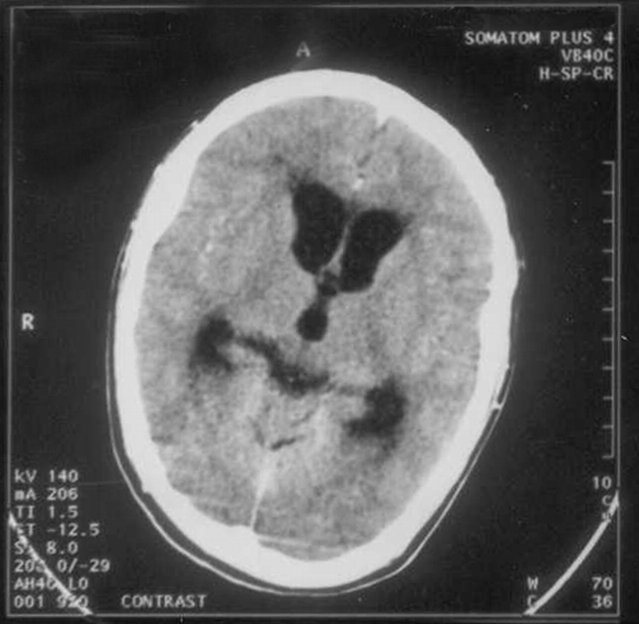
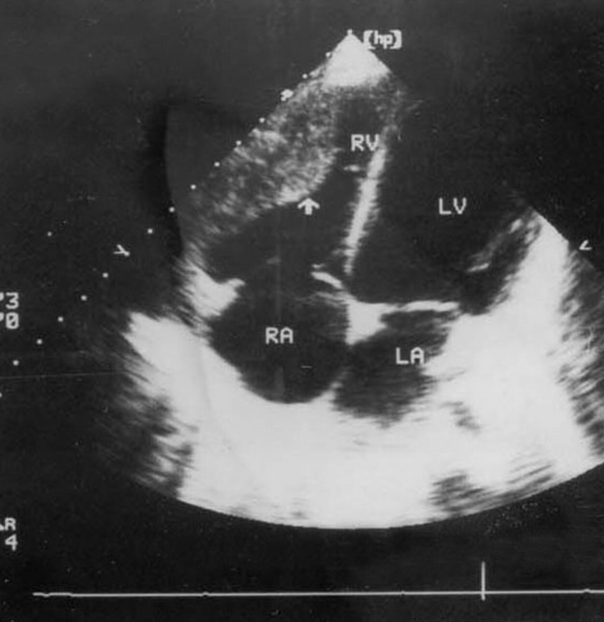
A 22-year-old man presented with a low-grade fever, headache and vomiting for 2 months, and 2 days of confusion and disorientation. He was diagnosed with tubercular meningitis after computed tomography (CT) of the cranium (Fig. 1) revealed mild hydrocephalus with dilatation of the third ventricle and the frontal and temporal horns of the lateral ventricles, in addition to mild periventricular ooze around the frontal horns of the lateral ventricles. A lumbar puncture was found to contain 0.64 g/L of protein and 710 white blood cells/mL with 74% lymphocytes. However, no acid-fast bacilli were seen on Ziehl-Neelsen staining. His chest radiograph was normal. He was admitted to hospital and started on antitubercular therapy (rifampicin, isoniazid, ethambutol and pyrazinamide). Although the results of his cardiac examination were unremarkable, an electrocardiogram performed routinely at the time of admission revealed multiple premature ventricular complexes. Two-dimensional echocardiography demonstrated an immobile, sessile mass along the right ventricular free wall (Fig. 2, arrow), which was also seen on the CT scan of the chest (Fig. 3). The patient declined an endomyocardial biopsy. Over the next 3 weeks, the patient became totally oriented and afebrile, and his headaches stopped. He was discharged home on the same 4-drug antitubercular therapy. Repeat echocardiography 6 months later revealed complete resolution of the mass, and his arrythmia was found to be resolved, thus suggesting a diagnosis of tuberculoma of the right ventricle.

Figure1
  • Download figure
  • Open in new tab
  • Download powerpoint

Figure 3. Photo: Images courtesy Dr. Amitabh Monga

Figure2
  • Download figure
  • Open in new tab
  • Download powerpoint

Figure 2. Photo: Images courtesy Dr. Amitabh Monga

Figure3
  • Download figure
  • Open in new tab
  • Download powerpoint

Figure 1. Photo: Images courtesy Dr. Amitabh Monga

Although tuberculosis (TB) primarily involves the lungs, 15%–20% of all cases are extrapulmonary, the most common sites being the lymph nodes, pleura, abdomen and central nervous system.1 Involvement of the heart (apart from the pericardium) is extremely rare and was first reported by Morgagni in 1761.2 Involvement of the myocardium occurs due to direct extension from the pericardium or spread from mediastinal lymph nodes or hematogenous dissemination. Three distinct histological forms of myocardial TB are recognized: diffuse infiltrating (the most common form, characterized microscopically by giant cells and lymphocytes), miliary (resulting from hematogenous spread) and nodular (characterized by central caseation).3,4,5 Ziehl-Neelsen staining of the endomyocardial biopsy specimens often fails to reveal acid-fast bacilli and definitive diagnosis rests on seeing typical histological changes.6 There are no specific features of a tuberculoma on a 2-dimensional echocardiogram or CT scan to distinguish it from other intracardiac masses.

Cardiac tuberculomas have been reported post mortem in fewer than 0.3% of all TB patients.2,7 Isolated cardiac tuberculomas are very rare.8 Single or multiple cardiac tuberculomas are most often observed in the right heart chambers, particularly in the wall of the right atrium. They are usually well circumscribed and sharply demarcated from the surrounding parenchyma. They may erode the underlying myocardium, resulting in ulcers that in turn cause thrombus formation and subsequent embolism. This may also result in hematogenous seeding and disseminated tuberculosis.9 Clinically, tuberculomas may be asymptomatic or may present with arrhythmias,5 complete heart block,8,10 congestive heart failure,11 superior vena caval obstruction,12 right ventricular outflow obstruction,13 aortic insufficiency14 and sudden cardiac death.15 In general, the prognosis is good and, as happened with our patient, resolution of the intracardiac mass and arrhythmias usually occurs after several months of antitubercular treatment.

Although cardiac tuberculomas are rare, they should be suspected in patients with arrhythmias or intracardiac masses who have been exposed to tuberculosis. With timely diagnosis, almost all these patients can be successfully treated with standard antitubercular therapy, though the exact duration of treatment that is required is still unclear.6

Amitabh Monga Anju Arora Ravinder Pal Singh Makkar Ajay K. Gupta Department of Internal Medicine Sitaram Bhartia Institute of Science and Research New Delhi, India

References

  1. 1.↵
    Fanning A. Tuberculosis: 6. Extrapulmonary disease. CMAJ 1999;160:1597-1603.
    OpenUrlFREE Full Text
  2. 2.↵
    Horn H, Saphir O. The involvement of myocardium in tuberculosis: a review of literature and a report of three cases. Am Rev Tuber 1935; 32: 492-504.
    OpenUrl
  3. 3.↵
    Gaultier Y, Alou A, Cenac A, Develoux M, Vetter JM. Tuberculome du coeur. Arch Mal Coeur 1987; 80:1413-6.
    OpenUrlPubMed
  4. 4.↵
    Baretti R, Eckel L, Beyersdorf F. Submitral left ventricular tuberculoma. Ann Thorac Surg 1995; 60 (1):81-2.
    OpenUrl
  5. 5.↵
    O'Neill PG, Rokey R, Greenberg S, Pacifico A. Resolution of ventricular tachycardia and endocardial tuberculoma following antituberculous therapy. Chest 1991;100(5):1467-9.
    OpenUrlCrossRefPubMed
  6. 6.↵
    Schnitzer R. Myocardial tuberculosis with paroxysmal ventricular tachycardia. Br Heart J 1947; 9:213-9.
    OpenUrlFREE Full Text
  7. 7.↵
    Gaultier Y, Alou A, Cenac A, Develoux M, Vetter JM. Tuberculoma of the heart. Contribution of echography. Apropos of a case. Arch Mal Coeur Vaiss 1987;80(9):1413-6.
    OpenUrlPubMed
  8. 8.↵
    Wren C, Stovin PG. Isolated interventricular septal tuberculoma causing complete heart block. Thorax 1982;37(2):149-50.
    OpenUrlFREE Full Text
  9. 9.↵
    Batra R, Trehan V, Salwan R, Krishan A, Nigam M, Malhotra V, et al. Antemortem diagnosis of cardiac tuberculoma. Indian Heart J 1998; 50(1): 87-9.
    OpenUrlPubMed
  10. 10.↵
    Kinare SG, Deshmukh MM. Complete atrioventricular block due to myocardial tuberculosis. Arch Pathol 1969;88:684-7.
    OpenUrlPubMed
  11. 11.↵
    Krishnaswami H, Cherian G. Right atrial tuberculoma: report of a case with complete recovery. Thorax 1984;39(7):550-1.
    OpenUrlFREE Full Text
  12. 12.↵
    Kapoor OP, Mascarenhas E, Rananaware MM, Gadgil RK. Tuberculoma of the heart. Report of 9 cases. Am Heart J 1973;86(3):334-40.
    OpenUrlCrossRefPubMed
  13. 13.↵
    Rawls WJ, ShufordWH, Logan WD, Hurst JW, Schlant RC. Right ventricular outflow tract obstruction produced by a myocardial abscess in a patient with tuberculosis. Am J Cardiol 1968;21: 738-45.
    OpenUrlCrossRefPubMed
  14. 14.↵
    Soyer R, Brunet A, Chevallier B, Leroy J, Morere M, Redonnet M. Tuberculous aortic insufficiency: report of a case with successful surgical treatment. J Thorac Cardiovasc Surg 1981; 82: 254-6.
    OpenUrlPubMed
  15. 15.↵
    Behr G, Palin HC, Temperley JM. Myocardial tuberculosis. BMJ 1977;1:951.
    OpenUrl
PreviousNext
Back to top

In this issue

CMAJ
Vol. 167, Issue 10
12 Nov 2002
  • Table of Contents
  • Index by author

Article tools

Respond to this article
Print
Download PDF
Article Alerts
To sign up for email alerts or to access your current email alerts, enter your email address below:
Email Article

Thank you for your interest in spreading the word on CMAJ.

NOTE: We only request your email address so that the person you are recommending the page to knows that you wanted them to see it, and that it is not junk mail. We do not capture any email address.

Enter multiple addresses on separate lines or separate them with commas.
A rare site for tuberculosis
(Your Name) has sent you a message from CMAJ
(Your Name) thought you would like to see the CMAJ web site.
CAPTCHA
This question is for testing whether or not you are a human visitor and to prevent automated spam submissions.
Citation Tools
A rare site for tuberculosis
Amitabh Monga, Anju Arora, Ravinder Pal Singh Makkar, Ajay K. Gupta
CMAJ Nov 2002, 167 (10) 1149-1150;

Citation Manager Formats

  • BibTeX
  • Bookends
  • EasyBib
  • EndNote (tagged)
  • EndNote 8 (xml)
  • Medlars
  • Mendeley
  • Papers
  • RefWorks Tagged
  • Ref Manager
  • RIS
  • Zotero
‍ Request Permissions
Share
A rare site for tuberculosis
Amitabh Monga, Anju Arora, Ravinder Pal Singh Makkar, Ajay K. Gupta
CMAJ Nov 2002, 167 (10) 1149-1150;
Digg logo Reddit logo Twitter logo Facebook logo Google logo Mendeley logo
  • Tweet Widget
  • Facebook Like
  • Google Plus One

Jump to section

  • Article
    • References
  • Figures & Tables
  • Responses
  • Metrics
  • PDF

Related Articles

  • No related articles found.
  • PubMed
  • Google Scholar

Cited By...

  • Unusual presentation of a young man with disseminated tuberculosis and right ventricular mass
  • Isolated right atrium tuberculoma causing complete heart block
  • Google Scholar

More in this TOC Section

  • The mystery of the broken bones
  • Elderly woman with rapidly growing, ulcerated pigmented lesion
  • Cat naps: an elderly woman with recurrent syncope
Show more Clinical Vistas

Similar Articles

Collections

  • Topics
    • Infectious diseases

Content

  • Current issue
  • Past issues
  • Collections
  • Sections
  • Blog
  • Podcasts
  • Alerts
  • RSS
  • Early releases

Information for

  • Advertisers
  • Authors
  • Reviewers
  • CMA Members
  • Media
  • Reprint requests
  • Subscribers

About

  • General Information
  • Journal staff
  • Editorial Board
  • Governance Council
  • Journal Oversight
  • Careers
  • Contact
  • Copyright and Permissions

Copyright 2021, CMA Joule Inc. or its licensors. All rights reserved. ISSN 1488-2329 (e) 0820-3946 (p)

All editorial matter in CMAJ represents the opinions of the authors and not necessarily those of the Canadian Medical Association or its subsidiaries.

To receive any of the resources on this site in an accessible format, please contact us at cmajgroup@cmaj.ca.

Powered by HighWire