
Main menu

  • Home
  • Content
    • Current issue
    • Past issues
    • Early releases
    • Collections
    • Sections
    • Blog
    • Infographics & illustrations
    • Podcasts
    • COVID-19 Articles
    • Obituary notices
  • Authors & Reviewers
    • Overview for authors
    • Submission guidelines
    • Submit a manuscript
    • Forms
    • Editorial process
    • Editorial policies
    • Peer review process
    • Publication fees
    • Reprint requests
    • Open access
    • Patient engagement
  • Members & Subscribers
    • Benefits for CMA Members
    • CPD Credits for Members
    • Subscribe to CMAJ Print
    • Subscription Prices
    • Obituary notices
  • Alerts
    • Email alerts
    • RSS
  • JAMC
    • À propos
    • Numéro en cours
    • Archives
    • Sections
    • Abonnement
    • Alertes
    • Trousse média 2023
    • Avis de décès
  • CMAJ JOURNALS
    • CMAJ Open
    • CJS
    • JAMC
    • JPN

User menu

Search

  • Advanced search
CMAJ
  • CMAJ JOURNALS
    • CMAJ Open
    • CJS
    • JAMC
    • JPN
CMAJ

Advanced Search

  • Home
  • Content
    • Current issue
    • Past issues
    • Early releases
    • Collections
    • Sections
    • Blog
    • Infographics & illustrations
    • Podcasts
    • COVID-19 Articles
    • Obituary notices
  • Authors & Reviewers
    • Overview for authors
    • Submission guidelines
    • Submit a manuscript
    • Forms
    • Editorial process
    • Editorial policies
    • Peer review process
    • Publication fees
    • Reprint requests
    • Open access
    • Patient engagement
  • Members & Subscribers
    • Benefits for CMA Members
    • CPD Credits for Members
    • Subscribe to CMAJ Print
    • Subscription Prices
    • Obituary notices
  • Alerts
    • Email alerts
    • RSS
  • JAMC
    • À propos
    • Numéro en cours
    • Archives
    • Sections
    • Abonnement
    • Alertes
    • Trousse média 2023
    • Avis de décès
  • Visit CMAJ on Facebook
  • Follow CMAJ on Twitter
  • Follow CMAJ on Pinterest
  • Follow CMAJ on Youtube
  • Follow CMAJ on Instagram
Review

Erythema ab igne: an old condition new again

Stephen Tan and Vince Bertucci
CMAJ January 11, 2000 162 (1) 77-78;
Stephen Tan
  • Find this author on Google Scholar
  • Find this author on PubMed
  • Search for this author on this site
Vince Bertucci
  • Find this author on Google Scholar
  • Find this author on PubMed
  • Search for this author on this site
  • Article
  • Responses
  • Metrics
  • PDF
Loading

Erythema ab igne (EAI) is a reticular, erythematous, hyperpigmented eruption that may occur after chronic exposure to moderate levels of infrared radiation in the form of heat.[1, 2] A case of EAI associated with the use of a space heater was reported by a 27-year-old healthy woman of South Asian background; she presented with a 3-month history of an asymptomatic rash on her lower legs, which was noticed 1 or 2 weeks after she began to use an electric space heater. The heater was placed approximately 30 cm away from the left side of her legs for 3-5 hours a day. The eruption began as reticulate erythematous patches on the lateral aspect of her left and the medial aspect of her right calf. She subsequently stopped using the heater and on presentation had nontender, reticulate, tan-brown patches with faint erythema in the above-mentioned areas. There was no evidence of telangiectasias, atrophy or nodules, and no systemic symptoms were reported.

Comment

EAI was once a common condition seen on the legs of those who stood or sat too close to a fire.[1, 3] Although the incidence of EAI has decreased since the introduction of central heating, it is still seen occasionally when localized areas of the body are repeatedly exposed to a heat source. The lesions seen by physicians now reflect the different sources of heat that are currently in use, and the distribution of EAI depends on the direction of the incident radiation, the contour of the skin and the interposition of clothing.1 EAI may result from the repeated application of hotwater bottles or heating pads used to treat chronic pain - backaches, for example, or pain associated with pancreatitis or peptic ulcer disease,[2, 4–7] as well as with primary or metastatic cancer.8 It may also appear on the skin of individuals who sit close to space heaters, wood-burning stoves or fireplaces,2 and it has been reported in association with exposure to a car heater9 and furniture with built-in heaters.[10, 11] EAI may be considered an occupational hazard for some people - the faces of silversmiths and jewelers and the arms of bakers, foundry workers and kitchen workers have been affected.[1, 2]

Following a single exposure to infrared radiation insufficient to cause a burn, a mild and transient reticular erythema may occur. Repeated exposure causes a more marked erythema with noticeable hyperpigmentation and, occasionally, superficial epidermal atrophy.1 The cumulative effects of exposure eventually resemble those of poikiloderma with erythema, hypo- and hyperpigmentation and reticulate telangiectasia, as well as diffuse hyperkeratosis.1 Subepidermal bullae may also occur.12 In severely affected individuals the reticular pattern is lost and a wide area of skin becomes pigmented and atrophic, with only the periphery showing the characteristic pattern.1 Apart from a slight burning sensation, the lesions of EAI are typically asymptomatic.13

Microscopic changes depend on the type of heat, the length of exposure and the area of the body involved.14 Histologically, EAI is similar to actinic keratoses,[2, 13, 15] with the epidermis showing squamous atypia.[13–17] There may also be an accumulation of dermal elastic tissue, which is an early sign of both UV radiation- and heat-induced skin damage.[2, 14] Squamous cell carcinoma and neuroendocrine carcinoma, also known as Merkel cell carcinoma, may arise in the lesions of EAI on rare occasions.[3, 18–20] The most common thermally induced cancer, squamous cell carcinoma,[3, 18] tends to occur after a long latent period of over 30 years.16 Squamous cell carcinoma in burn scars often begins as a chronic ulcer, which slowly enlarges and tends not to heal. Although these carcinomas tend to be of low to intermediate grade histologically, they may also be aggressive, with metastases and a poor prognosis reported in over 30% of cases.21 Neuroendocrine carcinoma is an aggressive neoplasm, characterized by local recurrence in approximately 30% of cases and up to 30% mortality.22 FIGURE

Figure

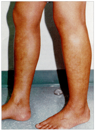
Fig. 1: Erythema ab igne on the legs. Note the reticulated, hyperpigmented patches on the lateral left and medial right calves.

The mainstay of treatment is to remove the source of infrared radiation immediately. Also, 5-fluorouracil cream has been reported to clear epithelial atypia.23 A biopsy should be done if there is any evidence of cutaneous malignancy, such as nodules or ulceration, within the EAI lesion. In many of the darker skin types the inflammatory process may lead to residual hypo- or hyperpigmentation; the patient in this case may experience hyperpigmentation that could persist for months to years, and this can be treated with topical tretinoin or hydroquinone.

The prognosis of EAI is excellent if the underlying infrared radiation source is removed early. However, if exposure to the infrared radiation is not discontinued, pigmentary abnormalities may persist and cutaneous atrophy or malignancy may ensue. The patient in this case presented with early stages of EAI and the heat source was promptly removed; it is likely, therefore, that her condition will resolve completely with time.

Physicians are well aware of the potential carcinogenic effects of cumulative UV radiation; the potential carcinogenicity of infrared radiation and the fact that it may enhance the aging and carcinogenic effects of UV exposure are not as well known, however.2 EAI lesions may be easily overlooked, but the diagnosis is more obvious if the patient's clinical history and the distribution and distinctive appearance of the cutaneous changes are considered. Because EAI may indicate an underlying malignancy that is causing chronic pain and because it can eventually develop into a carcinoma, it is important that the condition be properly diagnosed and followed.

Competing interests: None declared.

Footnotes

  • This article has been peer reviewed.

    Reprint requests to: Dr. Vince Bertucci, Division of Dermatology, Women's College Campus, Sunnybrook & Women's College Health Sciences Centre, 76 Grenville St., Toronto ON M5S 1B2.

References

  1. 1.↵
    Kennedy CTC. Reactions to mechanical and thermal injury. In: Champion RH, Burton JL, Ebling FJG, editors. Textbook of dermatology. 5th ed. Oxford: Blackwell Scientific Publications; 1992. p. 777-832.
  2. 2.↵
    Kligman LH, Kligman AM. Reflections on heat. Br J Dermatol 1984;110:369-75.
    OpenUrlCrossRefPubMed
  3. 3.↵
    Cross F. On a turf (peat) fire cancer: malignant change superimposed on erythema ab igne. Proc R Soc Med 1967;60:1307-8.
    OpenUrlFREE Full Text
  4. 4.↵
    Page EH, Shear NH. Temperature-dependent skin disorders. J Am Acad Dermatol 1988;18:1003-19.
    OpenUrlPubMed
  5. 5.
    Halliday CE, Goka AK, Farthing MJ. Erythema ab igne: a sign of organic disease [letter]. J R Soc Med 1986;79:249-50.
    OpenUrlFREE Full Text
  6. 6.
    Mok DW, Blumgart LH. Erythema ab igne in chronic pancreatic pain: a diagnostic sign. J R Soc Med 1984;77:299-301.
    OpenUrlAbstract/FREE Full Text
  7. 7.↵
    Butler ML. Erythema ab igne, a sign of pancreatic disease. Am J Gastroenterol 1977;67:77-9.
    OpenUrlPubMed
  8. 8.↵
    Ashby M. Erythema ab igne in cancer patients. J R Soc Med 1985;78:925-7.
    OpenUrlAbstract/FREE Full Text
  9. 9.↵
    Helm TN, Spigel GT, Helm KF. Erythema ab igne caused by a car heater. Cutis 1997;59:81-2.
    OpenUrlPubMed
  10. 10.↵
    Meffert JJ, Davis BM. Furniture-induced erythema ab igne. J Am Acad Dermatol 1996;34:516-7.
    OpenUrlCrossRefPubMed
  11. 11.↵
    Dvoretzky I, Silverman NR. Reticular erythema of the lower back. Arch Dermatol 1991;127:405-9.
  12. 12.↵
    Flanagan N, Watson R, Sweeney E, Barnes L. Bullous erythema ab igne [letter]. Br J Dermatol 1996;134:1159-60.
    OpenUrl
  13. 13.↵
    Shahrand P, Marks R. The wages of warmth: changes in erythema ab igne. Br J Dermatol 1977;97:179-86.
    OpenUrlCrossRefPubMed
  14. 14.↵
    Johnson WC, Butterworth T. Erythema ab igne elastosis. Arch Dermatol 1971;104:128-31.
    OpenUrlCrossRefPubMed
  15. 15.↵
    Arrington JH, Lockman DS. Thermal keratoses and squamous cell carcinoma in situ associated with erythema ab igne. Arch Dermatol 1979;115:1226-8.
    OpenUrlCrossRefPubMed
  16. 16.↵
    Galvin, SA, Buchness MR. Rectangular reticulate patches on the pretibial areas. Arch Dermatol 1990;126:386.
    OpenUrlCrossRefPubMed
  17. 17.↵
    Finlayson GR, Sams WM, Smith JG. Erythema ab igne: a histopathological study. J Invest Dermatol 1966;46:104-8.
    OpenUrlPubMed
  18. 18.↵
    Peterkin GA. Malignant change in erythema ab igne. BMJ 1955;2:1599-600.
  19. 19.
    Jones, CS, Tyring SK, Lee PC, Fine JD. Development of neuroendocrine (Merkel cell) carcinoma mixed with squamous cell carcinoma in erythema ab igne. Arch Dermatol 1988;124:110-3.
    OpenUrlCrossRefPubMed
  20. 20.↵
    Hewitt JB, Sherif A, Kerr KM, Stankler L. Merkel cell and squamous cell carcinomas arising in erythema ab igne [letter]. Br J Dermatol 1993;128:591-2.
    OpenUrlCrossRefPubMed
  21. 21.↵
    Kaplau RP. Cancer complicating chronic ulcerative and scarifying mucocutaneous disorders. Adv Dermatol 1987;2:19-46.
    OpenUrlPubMed
  22. 22.↵
    Sibley RK, Dehmer LP, Rosai J. Primary neuroendocrine (Merkel cell?) carcinoma of the skin 1. A clinicopathologic and ultrastructural study of 43 cases. Am J Surg Pathol 1985;9:95-108.
    OpenUrlCrossRefPubMed
  23. 23.↵
    Sahl WJ, Taira JW. Erythema ab igne: treatment with 5-fluorouracil cream. J Am Acad Dermatol 1992;27:109-10.
    OpenUrlPubMed
PreviousNext
Back to top

In this issue

CMAJ
Vol. 162, Issue 1
11 Jan 2000
  • Table of Contents
  • Index by author

Article tools

Respond to this article
Print
Download PDF
Article Alerts
To sign up for email alerts or to access your current email alerts, enter your email address below:
Email Article

Thank you for your interest in spreading the word on CMAJ.

NOTE: We only request your email address so that the person you are recommending the page to knows that you wanted them to see it, and that it is not junk mail. We do not capture any email address.

Enter multiple addresses on separate lines or separate them with commas.
Erythema ab igne: an old condition new again
(Your Name) has sent you a message from CMAJ
(Your Name) thought you would like to see the CMAJ web site.
CAPTCHA
This question is for testing whether or not you are a human visitor and to prevent automated spam submissions.
Citation Tools
Erythema ab igne: an old condition new again
Stephen Tan, Vince Bertucci
CMAJ Jan 2000, 162 (1) 77-78;

Citation Manager Formats

  • BibTeX
  • Bookends
  • EasyBib
  • EndNote (tagged)
  • EndNote 8 (xml)
  • Medlars
  • Mendeley
  • Papers
  • RefWorks Tagged
  • Ref Manager
  • RIS
  • Zotero
‍ Request Permissions
Share
Erythema ab igne: an old condition new again
Stephen Tan, Vince Bertucci
CMAJ Jan 2000, 162 (1) 77-78;
Digg logo Reddit logo Twitter logo Facebook logo Google logo Mendeley logo
  • Tweet Widget
  • Facebook Like

Jump to section

  • Article
    • Comment
    • Footnotes
    • References
  • Responses
  • Metrics
  • PDF

Related Articles

  • No related articles found.
  • PubMed
  • Google Scholar

Cited By...

  • A Hyperpigmented Reticular Rash in a Patient on Peritoneal Dialysis
  • Academic branding: erythema ab igne and use of laptop computers
  • Laptop Computer-Induced Erythema ab Igne in a Child and Review of the Literature
  • Answer: Can you identify this condition?
  • Erythema ab igne
  • Google Scholar

More in this TOC Section

  • Diagnosis and management of endometriosis
  • Diagnosis and management of patients with polyneuropathy
  • Pharmacologic prevention of migraine
Show more Review

Similar Articles

Collections

  • Topics
    • Dermatology
    • Environmental issues


View Latest Classified Ads

Content

  • Current issue
  • Past issues
  • Collections
  • Sections
  • Blog
  • Podcasts
  • Alerts
  • RSS
  • Early releases

Information for

  • Advertisers
  • Authors
  • Reviewers
  • CMA Members
  • CPD credits
  • Media
  • Reprint requests
  • Subscribers

About

  • General Information
  • Journal staff
  • Editorial Board
  • Advisory Panels
  • Governance Council
  • Journal Oversight
  • Careers
  • Contact
  • Copyright and Permissions
  • Accessibiity
  • CMA Civility Standards
CMAJ Group

Copyright 2023, CMA Impact Inc. or its licensors. All rights reserved. ISSN 1488-2329 (e) 0820-3946 (p)

All editorial matter in CMAJ represents the opinions of the authors and not necessarily those of the Canadian Medical Association or its subsidiaries.

To receive any of these resources in an accessible format, please contact us at CMAJ Group, 500-1410 Blair Towers Place, Ottawa ON, K1J 9B9; p: 1-888-855-2555; e: cmajgroup@cmaj.ca

Powered by HighWire