


- © 2007 Canadian Medical Association or its licensors
What's your call?
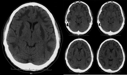
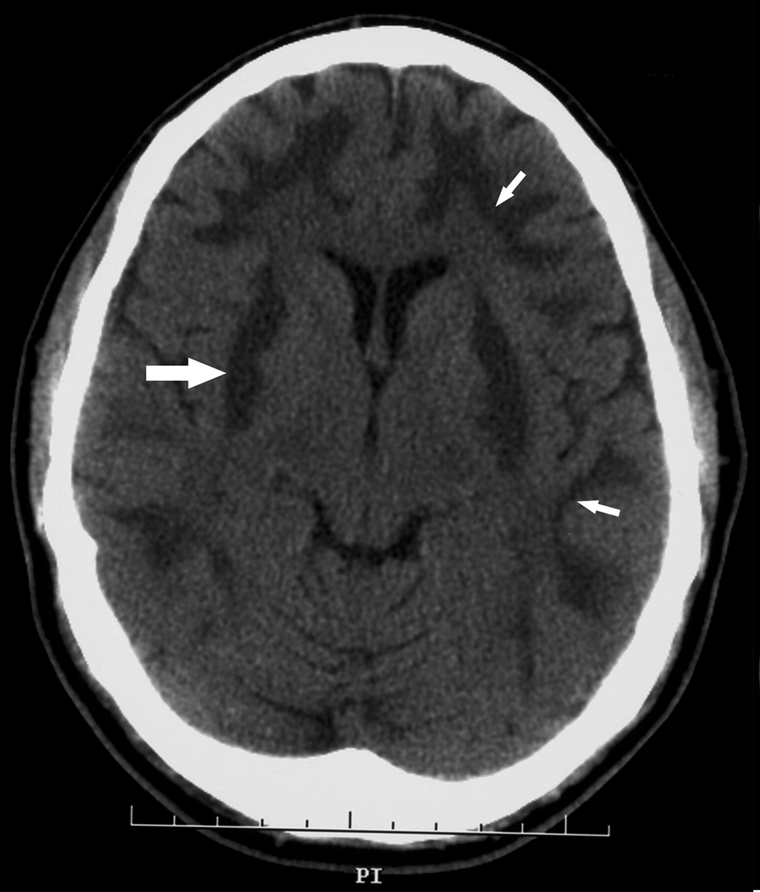
Figure. Head CT scans of an unconscious 42-year-old man, taken 48 hours after admission to hospital after an attempted suicide.
The patient had attempted suicide by consuming methanol. On admission, his methanol level was 46 (normal < 2) mmol/L. He was unconscious, with generalized rigidity, exaggerated muscle stretch reflexes, and bilateral clonus and extensor plantar responses. CT scans of the brain obtained 48 hours after admission to hospital showed symmetric areas of low attenuation in the peripheral white matter of both cerebral hemispheres bordered by the grey-white junction, involving both frontal, parietal occipital and temporal lobes (Fig. 1, small arrows). Symmetric hypointensities were also noted in both lentiform nuclei, centred within the putamen and extending into the external capsules (Fig. 1, bold arrow).

Fig. 1: CT scan showing symmetrical areas of low attenuation in the peripheral white matter (small arrows) and symmetrical hypointesities in both lentiform nuclei (bold arrow).
Mechanical ventilation, hemodialysis and intravenous ethanol therapy were begun, with no significant improvement in clinical status. Nine days after admission, the patient died of respiratory failure after his family decided to withdraw ventilatory support.
Methanol has a toxic effect on the central nervous system, especially the optic nerves and the basal ganglia. Symmetric involvement of the basal ganglia is the most characteristic radiologic feature of methanol poisoning.1 However, certain other conditions must be kept in mind in the differential diagnosis, such as carbon monoxide intoxication,2 striatonigral degeneration, anoxic stroke and Wilson's disease.3 Although bilateral involvement of the basal ganglia on CT scans is a characteristic finding of methanol intoxication, it is infrequently seen in clinical practice.
Footnotes
-
Competing interests: None declared.
In this issue
Article tools


Jump to section
Related Articles
Cited By...
- No citing articles found.
More in this TOC Section
Similar Articles

